
By evening, your legs feel heavy, your ankles look puffy, and a cluster of bulging veins seems more noticeable than it did that morning. Or your legs look almost normal, yet they ache after standing. You may be wondering whether stockings are enough, whether a procedure would help, or why a wound near your ankle won't close.
Venous reflux disease treatment starts with finding where blood is flowing backward, not choosing which visible veins to remove. The right plan connects your symptoms, ultrasound findings, and overall circulation. If you have an open ulcer, it also needs to account for arterial blood flow, infection, and the actual cause of the wound.
Table of Contents
- Treatment Options for Venous Reflux at a Glance
- How Doctors Confirm Reflux Before Choosing Treatment
- Compression and Lifestyle Care That Still Matters
- How Endovenous Procedures Close the Source of Reflux
- Why Veins Come Back and What Follow-Up Prevents
- Deciding Treatment Timing When a Leg Ulcer Is Open
- Wound-Focused Management for Open Venous Ulcers
- Confirm a Specialist Evaluation Before You Commit
Treatment Options for Venous Reflux at a Glance
Healthy leg veins contain one-way valves that help blood travel upward toward the heart. The valves close between pushes from the calf muscles, limiting backward flow. When a valve fails to close properly, blood can leak backward and pool below it. That flow is venous reflux.
Reflux can contribute to aching, heaviness, fatigue, swelling, bulging veins, and changes in the skin around the lower leg. These symptoms warrant evaluation, though they do not establish reflux as the only cause. A swollen ankle, for example, may have a different explanation from a nearby bulging vein.
A truncal vein is a main superficial vein that collects blood from smaller branches. When it refluxes, treating only visible branches may leave the source of pressure untreated.
Match the treatment to the problem
For confirmed varicose veins with truncal reflux, the NICE varicose-vein guideline recommends endothermal ablation first, ultrasound-guided foam sclerotherapy when endothermal ablation is unsuitable, and surgery when foam is unsuitable. Guidelines set an order of options, and a given patient may use only some of them.
Venous reflux treatment options by use, setting and outcome
| Option | Typical use | Setting | Key outcome | Refer when |
|---|---|---|---|---|
| Compression and leg elevation | Mild symptoms, swelling, and recurrence prevention after ulcer healing | Daily use at home | Helps control heaviness and swelling; ongoing compression supports recurrence prevention | Symptoms persist despite consistent use |
| Endovenous laser ablation | Truncal great saphenous reflux | Outpatient, local anaesthetic | Closes the refluxing trunk from within | Suitability needs assessment, including anatomy and comorbidities |
| Radiofrequency ablation | Truncal reflux suitable for heat-based closure | Outpatient, local anaesthetic | Heat seals the unhealthy vein and redirects blood through healthier pathways | Anatomy or comorbidity may make heat-based treatment unsuitable |
| Ultrasound-guided foam sclerotherapy | Reflux unsuitable for thermal ablation, and selected tributaries | Outpatient | Closes the treated segment; additional treatment may be needed | Symptoms or reflux persist after treatment |
| Ambulatory phlebectomy | Bulging surface varicosities | Outpatient, through small incisions | Removes visible branches but doesn't independently correct an untreated refluxing trunk | Visible veins return, symptoms persist, or an ulcer is present |
| Surgical vein removal | When ablation and foam are unsuitable | Hospital or day-case theatre | Removes the selected unhealthy vein | Minimally invasive options are unsuitable or unsuccessful |
| Wound-focused care | Active venous leg ulcer | Wound clinic | Supports healing while addressing circulation and compression needs | A lower-leg wound remains open or deteriorates |
Each option addresses a different part of the problem. Compression and elevation can help control pressure-related symptoms, but they do not repair a valve. Ablation closes a selected abnormal pathway, while phlebectomy removes surface branches. Wound-focused care supports an open ulcer and its circulation and compression needs.
More than one approach may be appropriate. A person with an ankle ulcer and superficial reflux may need wound care, compression, and a reflux-directed procedure together. Someone whose symptoms are tolerable and who has no skin damage may start with conservative management. For an open ulcer, treatment timing also depends on checking arterial circulation, infection, and whether the wound is venous in origin.
Practical rule: Use the table to identify which problem an option addresses, then confirm whether it fits the person's symptoms and findings. It is a guide to the choices, not a decision on its own.
How Doctors Confirm Reflux Before Choosing Treatment
The veins you can see aren't always the veins causing your symptoms. A prominent surface branch may connect to a refluxing trunk elsewhere, while clinically important reflux may be present without dramatic bulging.
Our consultation and duplex ultrasound help identify where abnormal flow occurs. We base the plan on the source of the problem, rather than appearance alone.
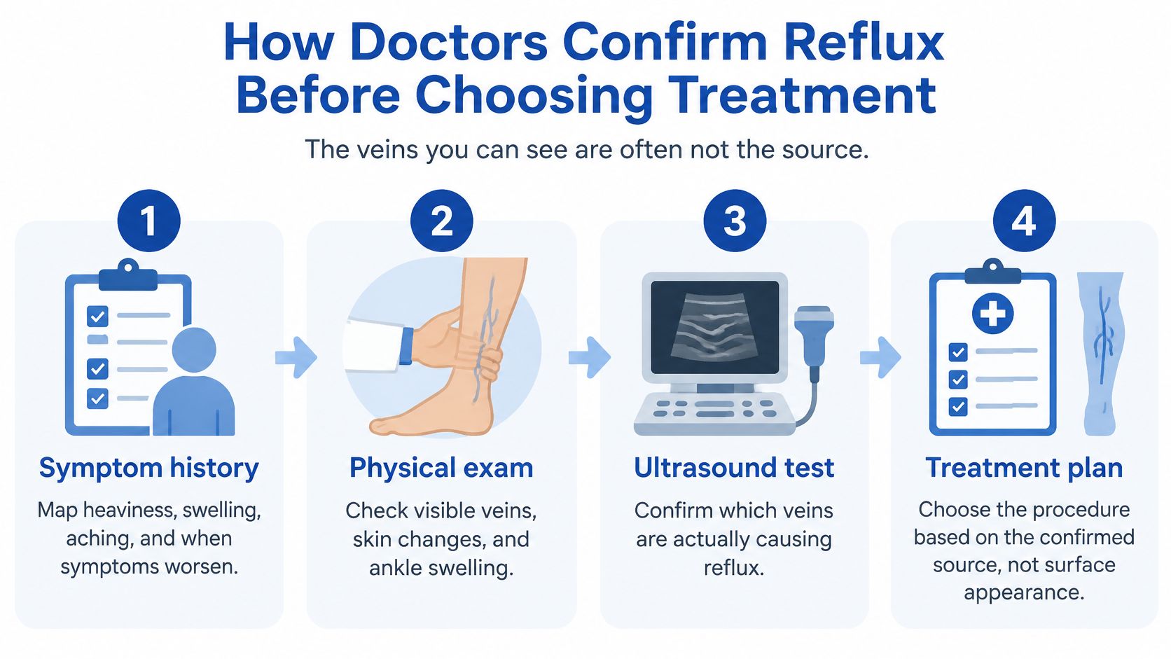
The diagnostic sequence
Review symptoms and history. Describe when discomfort starts, whether elevation helps, and how swelling changes during the day. Prior ulcers, vein procedures, clots, and family history help the clinician interpret what's happening now. A common mistake is discussing the visible veins but leaving out an old ankle wound.
Examine the legs and skin. The clinician checks swelling, vein distribution, pigmentation, eczema-like changes, and healed or open wounds. The CEAP classification, a framework describing clinical findings, cause, anatomy, and vein function, can help document severity. Appearance alone can't identify the full flow problem.
Map flow with duplex ultrasound. Testing commonly includes upright assessment, when feasible, to examine reflux under conditions that challenge the valves. The map can include the great and small saphenous veins, connecting perforator veins, and the deep system. Our guide to ultrasound for chronic venous insufficiency explains how this test helps distinguish an abnormal source from a visible branch.
Check arterial circulation when indicated. An ulcer, concerning pain, or examination findings may call for an ankle-brachial index, which compares ankle and arm blood pressures, or another arterial test. This is the safety check patients can overlook: finding reflux doesn't establish that strong compression is safe.
Assess any wound for infection and other causes. The clinician looks for spreading redness, warmth, increasing pain, drainage changes, and systemic illness. A swab isn't automatic for every open wound; sampling is guided by clinical findings and whether it will help treatment.
Combine the findings into a written plan. The plan should identify the abnormal pathway, explain how it relates to your symptoms, and name the appropriate treatment class. If symptoms and ultrasound don't match, the clinician should consider another explanation rather than treating a vein because it is enlarged.
What you should understand before leaving
You should know which veins reflux, whether deep or arterial disease complicates the picture, and what the proposed treatment targets. You don't need to memorize the scan report, but you should be able to describe the reason for treatment in plain language.
For example, “The main superficial vein is leaking backward and feeding these branches” is a more useful explanation than “You have bad veins.”
Compression and Lifestyle Care That Still Matters
Compression is foundational care, and using it well does not mean treatment has failed. Stockings apply external pressure to limit swelling and support venous return. They can be part of care while a treatment plan is developed, alongside a procedure, or for ongoing management when that approach suits the patient's condition and goals.
A vein consultation and duplex ultrasound help identify where abnormal reflux is occurring and which treatment is most appropriate. If symptoms persist, or compression, elevation and activity changes do not provide enough relief, a minimally invasive procedure may be considered. Treatment recommendations are personalized, with the source of the problem guiding the plan rather than the veins visible at the skin's surface.
Build a routine you can sustain
Use clinician-selected compression. Below-knee graduated stockings are a common option. Their strength and design should reflect your circulation, skin condition, and ability to put them on. Wear them according to the prescribed schedule, ideally before swelling becomes pronounced.
Rest with support. When practical, raise your legs comfortably above heart level. Support the lower legs without pressing a chair edge into the calves. The position should ease discomfort and leave your feet comfortable.
Activate the calf muscles. Walk within your mobility limits. While seated, ankle pumps and gentle heel movements help keep the calves moving.
Break up prolonged stillness. Alternate sitting, standing, and walking when possible. If weight management is relevant, a workable plan can be part of care.
For ulcer-related disease, the NICE recommendations define a suspected venous leg ulcer as a below-knee skin break that has not healed within two weeks and recommend vascular assessment. The guidance also supports below-knee graduated compression after healing to reduce recurrence risk.
Know when to adjust the plan
When symptoms are mild, daily function is acceptable, and there is no open ulcer or concerning skin damage, conservative care can be a reasonable approach. Returning swelling, persistent aching, or new skin changes are reasons to seek reassessment. An evaluation can help determine the severity of chronic venous disease and whether the current approach still fits.
Stockings that cause pain need a fit and safety check. Rolling fabric, incorrect sizing, excessive pressure, or inadequate arterial circulation can contribute to discomfort. Ask about refitting, a different prescribed compression level, an open-toe design, an application aid, or an adjustable wrap. Guidance on compression stockings for chronic venous insufficiency explains their role in symptom management.
Marked pain, numbness, or a change in foot colour or temperature calls for prompt clinical advice. Compression should be comfortable enough to use as directed, and the plan should be reviewed when symptoms or skin changes continue.
How Endovenous Procedures Close the Source of Reflux
A patient may have a refluxing great saphenous vein while the deep veins continue to carry blood toward the heart. Closing the faulty superficial vein can redirect blood through healthier routes, helping reduce venous pressure and symptoms. The target is the source of reflux, which may not be the vein that looks most prominent at the skin.
Endovenous treatment works from inside the vein. Laser and radiofrequency procedures use heat to seal the treated segment; foam sclerotherapy uses an injected agent. Traditional stripping removes the vein through incisions, rather than closing it from within.
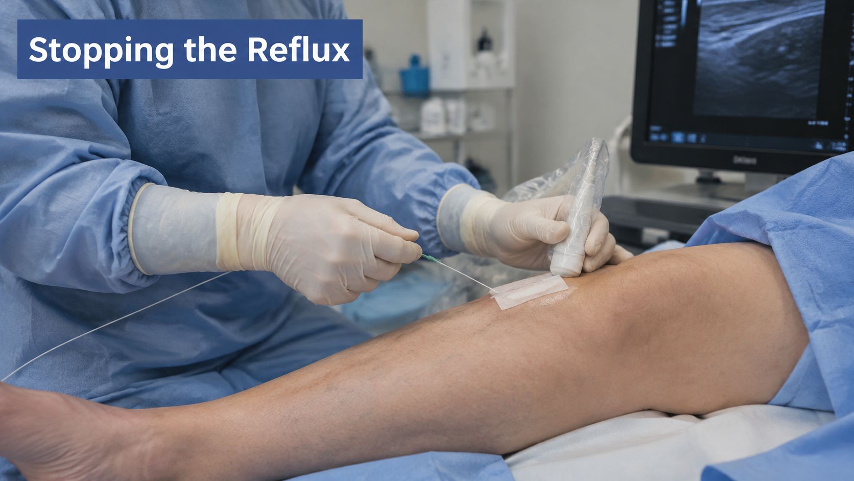
What thermal ablation involves
- Ultrasound-guided access. The clinician enters the selected vein through a small access point and positions the treatment catheter.
- Local anaesthetic around the vein. Fluid placed around the vessel numbs the area and helps protect nearby tissues during heat treatment.
- Controlled energy delivery. Laser or radiofrequency energy treats the intended segment as the device is withdrawn.
- Procedure-specific aftercare. Compression may be applied, and light walking is generally encouraged according to the discharge plan.
Foam treatment follows a different process and does not use a heating catheter. The choice depends on the target vein's anatomy and the treatment goal.
What closure results actually mean
The Cochrane review comparing endovenous treatment with surgery reported lower odds of persistent reflux or recanalization after EVLA at six weeks and lower odds of recurrent reflux at one year. It found no statistically clear difference in newly visible varicose veins at one year.
Cochrane outcomes after EVLA compared with surgery, review results
| Outcome | Comparison | Odds ratio | 95% CI | Participants |
|---|---|---|---|---|
| Persistent reflux or recanalization at six weeks | EVLA vs surgery | 0.07 | 0.02–0.22 | 289 |
| Recurrent reflux at one year | EVLA vs surgery | 0.24 | 0.07–0.77 | 119 |
| Clinical recurrence (new visible veins) at one year | EVLA vs surgery | 0.54 | 0.17–1.75 | 99 |
An odds ratio compares the odds of an outcome between groups. The results favour EVLA for the ultrasound-defined reflux outcomes, but they do not predict an individual patient's outcome. For visible-vein recurrence, the uncertainty is too broad to establish a clear difference.
Closing the intended vein and preventing every future visible vein are separate goals. Discuss symptom improvement, remaining branches, and possible additional treatment separately.
Surgical removal remains an option when less invasive approaches are unsuitable. It involves incisions and may require more recovery planning than an outpatient catheter procedure. Neither approach is risk-free. Consent should cover relevant possibilities, including bruising, discomfort, clot-related complications, nerve irritation, and alternatives suited to the patient's anatomy.
Why Veins Come Back and What Follow-Up Prevents
A successfully closed vein doesn't mean the underlying chronic disease has ended. A person can have an excellent technical result and still need ongoing management for deep venous reflux, obstruction, lymphatic dysfunction, or limited calf-muscle activity.
Persistent swelling also doesn't automatically mean the treated vein reopened. If fluid clearance remains impaired, repeating superficial treatment without reassessment may miss the underlying problem.
The clinical review on compression therapy for chronic venous disease and venous ulceration describes factors associated with treatment failure or recurrence, including older age, obesity, deep reflux, arterial insufficiency, poor compression adherence, large or longstanding ulcers, and repeated ulcer episodes. These factors help explain why follow-up intensity should reflect the patient's overall disease, not just the procedure performed.
Separate recovery from long-term surveillance
Your immediate discharge plan may include compression, temporary limits on strenuous exercise, and specific bathing or access-site instructions. These directions vary by procedure, so don't substitute someone else's recovery schedule for your own.
Longer-term review asks different questions:
- Has the target stayed closed? Duplex reassessment can investigate residual or recurrent reflux when indicated.
- Are symptoms improving as expected? Persistent aching or swelling may require a broader assessment.
- Can you use the prescribed compression? Difficulty applying stockings is a treatment barrier to solve, not a reason to label someone uncooperative.
- Is the skin staying healthy? New irritation, discolouration, or breakdown may call for a revised prevention plan.
An office may describe a scheduled review as a recall appointment. TOOLii's explanation of what a recall appointment means can help clarify the scheduling term, although the purpose and timing of your vascular review come from your own clinical plan.
Don't assume all swelling is recurrent reflux
Lymphedema, reduced mobility, mixed vascular disease, and other medical causes can contribute to ongoing swelling. The distribution of swelling, skin findings, examination, and imaging help determine whether another vein procedure would make sense.
A follow-up visit is useful even when no additional procedure is needed. It can identify a compression problem, a different cause of swelling, or early skin breakdown.
Before discharge, clarify how long compression is intended, which activities are temporarily restricted, and who will reassess symptoms that don't improve. If swelling continues, ask whether a lymphatic assessment belongs in the plan.
Deciding Treatment Timing When a Leg Ulcer Is Open
An open leg ulcer can raise a practical question: should reflux treatment wait until the wound closes? For a suitable patient with treatable superficial reflux, an ulcer does not by itself rule out intervention. Treating abnormal blood flow alongside compression and wound care may support healing.
The wound's location can offer a clue, but it cannot identify the cause on its own. An ankle ulcer may be venous, arterial, diabetic, pressure-related, or have mixed causes. The treatment decision depends on checking both the wound and the circulation.
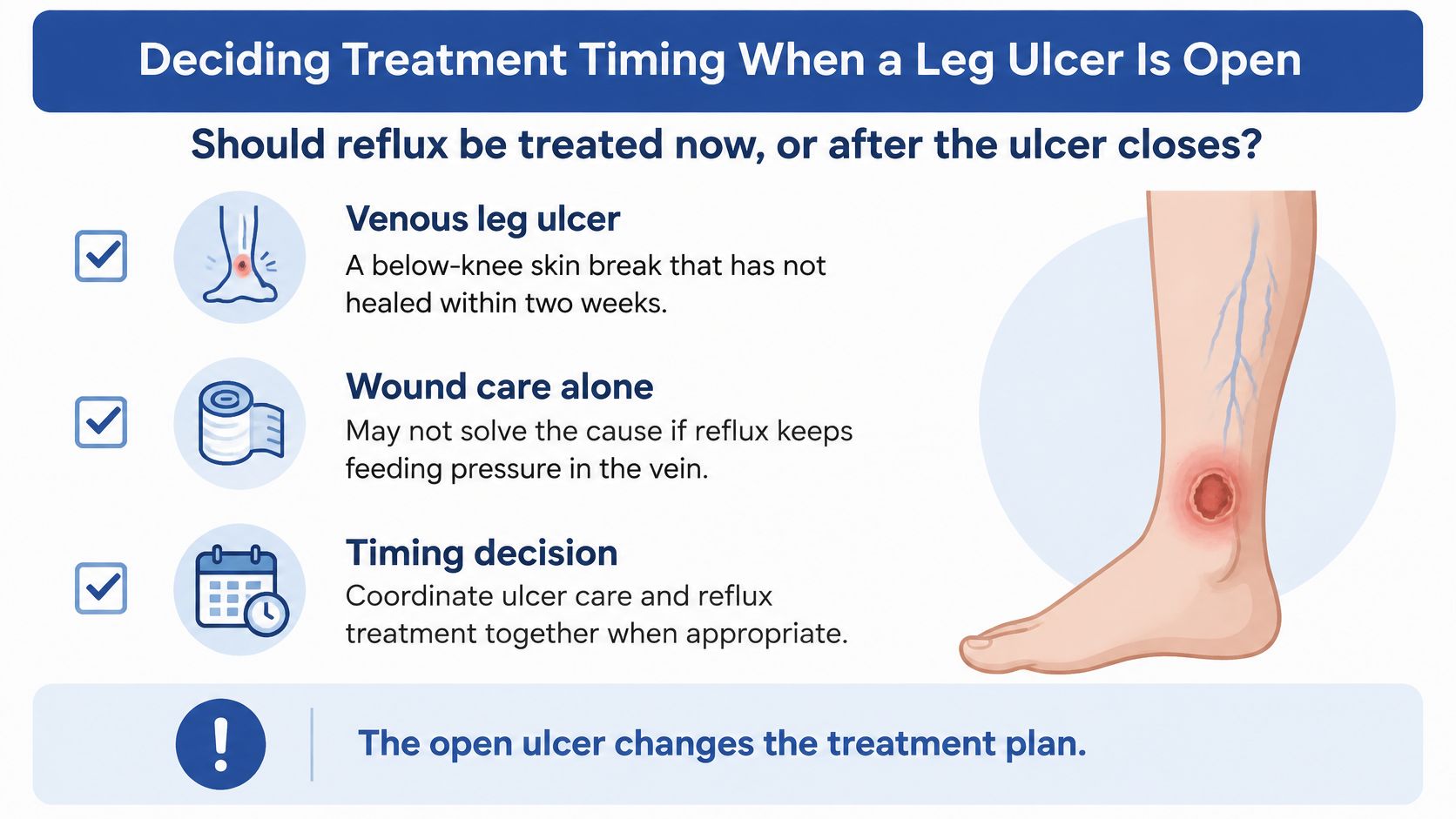
What early treatment can change
A randomized trial of 450 patients, described in the British Journal of Dermatology article, compared early endovenous ablation plus compression with deferred treatment. The reported outcomes were:
Trial outcomes for early ablation plus compression in open ulcers
| Measure | Early ablation plus compression | Comparator arm |
|---|---|---|
| Median healing time | 56 days | 82 days |
| Healing at 24 weeks | 85.6% | 76.3% |
| Hazard ratio for healing (ablation vs deferred) | 1.38 | Deferred treatment |
These results show why treatment timing may matter for selected patients with superficial reflux. Ablation addresses the abnormal vein, while compression and wound care continue to support healing. The trial outcomes do not mean that ablation replaces either part of that care.
Checks that determine the timing
- Arterial supply. Pulse checks, an ankle-brachial index, or another suitable arterial assessment help determine whether compression is safe and whether arterial treatment should take priority.
- Infection status. Spreading inflammation, increasing pain, purulent drainage, or systemic illness can change immediate care.
- Ulcer size and duration. A large or longstanding wound calls for realistic healing expectations and coordination between vascular and wound care.
- Deep venous findings. Deep reflux or obstruction may affect expected results and call for further vascular evaluation.
- Diabetic or pressure-related injury. Neuropathy, repeated pressure, and footwear or positioning issues need to be addressed as part of the plan.
- Mixed causes. More than one factor may be slowing repair. Finding superficial reflux does not resolve other contributors.
The trial evidence applies to selected patients with superficial reflux, not every wound that has failed to heal. If inadequate arterial supply or ongoing pressure is overlooked, treating a vein that is not the main driver may delay appropriate care.
Timing principle: Assess the wound and circulation together. An open ulcer alone need not delay suitable reflux treatment, while the cause of the wound must be established before choosing an intervention.
A coordinated plan should specify who manages dressings and compression, and how the vascular and wound teams will assess progress.
Wound-Focused Management for Open Venous Ulcers
An ulcer may remain open even when the visible veins seem unremarkable. Care should address the wound and the circulation feeding it, with reflux treated as a possible source of ongoing pressure rather than judging the problem by appearance alone.
Follow a coordinated wound plan
Confirm wound type and arterial safety. Check pulses and arrange arterial testing when indicated. If arterial disease is also present, it can change which compression system and pressure are appropriate.
Clean and debride selectively. The wound team removes material that interferes with healing when appropriate and chooses dressings based on drainage, surrounding skin, and wound depth. For example, reduced blood flow may affect whether and how a wound is debrided.
Assess possible infection. A clinician looks at the wound and surrounding skin to decide whether sampling or infection treatment is appropriate. Spreading inflammation, worsening pain, purulent drainage, or systemic illness calls for urgent attention.
Use suitable compression. Once arterial status permits, apply the clinician-selected system and check its fit, tolerance, and effect on the skin. New pain, coldness, colour changes, or unexpected swelling warrants reassessment.
Track healing over time. Record wound dimensions and appearance at follow-up. If an ulcer stops shrinking after six weeks despite correct dressings, stalled healing should prompt a fresh look at circulation, residual reflux, pressure, diabetes-related injury, and other possible causes.
Match vein treatment to the whole wound plan. Closing or removing an unhealthy vein may help reduce venous pressure, but the care plan also needs to address wound management and circulation. The choice of intervention depends on the patient's findings and suitability.
The guide to chronic venous insufficiency ulcers explains more about ulcer-related care. Diabetic and pressure-related wounds need care directed at those causes, even when venous disease is present too.
Bring previous wound measurements, dressing names, photographs showing changes, and records of prior infection treatment. These details help the care team see whether the wound is improving, has stalled, or has worsened.
Confirm a Specialist Evaluation Before You Commit
Before agreeing to a procedure, prepare a short account of what bothers you and what you want treatment to improve. Include when symptoms began, how they affect walking or work, any current or previous ulcers, past vein procedures, and a history of clots or venous disease in the family.
Our evaluation helps determine disease severity, and recommendations are personalized to your condition and goals. Procedure selection rests on vein size and location, ultrasound findings, symptoms, and overall vascular health, not a photograph or a preferred device.
Bring questions that test the plan
- Which veins are refluxing? Ask to have the ultrasound findings explained in ordinary language.
- What does the proposed treatment target? Distinguish treatment of the abnormal source from removal of visible branches.
- What improvement is realistic? Separate relief of aching, reduction of swelling, wound healing, and cosmetic change.
- What happens afterward? Clarify compression, recovery instructions, and how persistent or returning symptoms will be assessed.
If you have a non-healing wound, request an evaluation that includes both circulation and wound assessment. A plan that addresses only the vein or only the dressing may leave an important cause untreated.
Recognize symptoms that shouldn't wait
Arrange prompt assessment for an open lower-leg wound, new skin breakdown, or worsening symptoms. Sudden painful swelling of one leg needs same-day assessment, because it shouldn't be assumed to be routine reflux.
Active bleeding also requires immediate attention: lie down, raise the leg, and apply firm direct pressure. Seek emergency help for heavy bleeding, bleeding that won't stop, chest pain, breathlessness, or severe illness; spreading wound redness with fever also warrants urgent care.
This week, gather your records and arrange a source-focused evaluation. The useful decision isn't whether to have treatment, but whether the proposed treatment matches your problem.
Vein and Wound Institute brings vein evaluation, duplex ultrasound, minimally invasive treatment, and specialized wound management together with coordinated follow-up. Visit Vein and Wound Institute to arrange an evaluation for persistent leg symptoms or a non-healing wound and discuss a plan based on your circulation, symptoms, and goals.