
In a United States study of healthcare workers, reflux was detected in at least one leg in 82.1% of participants, whereas venous obstruction was found in only 0.2%, according to ultrasound-based population research. That gap changes how you should read a chronic venous insufficiency ultrasound report. Finding backward flow is common. Finding a blocked vein is not. Neither finding, on its own, tells you how severe your symptoms are or whether a wound can heal safely with compression.
A good report answers narrower, more useful questions. Which vein is leaking, where does the leak start, how far does it extend, and is anything blocked? It doesn't assess whether your arteries deliver enough blood to support healing. That distinction matters before anyone recommends a procedure, compression, or wound treatment, and it's the distinction many patient guides skip.
Table of Contents
- What Duplex Ultrasound Shows and What the Numbers Mean
- Reflux, Obstruction and How Common Each Finding Is
- Why Duplex Ultrasound Is the First Test for Venous Disease
- How to Prepare for and Undergo the Scan
- What Ultrasound Cannot Tell You
- From Duplex Map to Treatment, Follow-Up and Next Steps
What Duplex Ultrasound Shows and What the Numbers Mean
| Finding | Ultrasound sign or threshold | What it means |
|---|---|---|
| Superficial vein reflux | Reversed flow longer than 0.5 seconds | Abnormal backward flow in a superficial vein or tributary |
| Perforator reflux | Reversed flow longer than 0.5 seconds | Abnormal flow between superficial and deep systems |
| Deep vein reflux (common femoral, femoral, popliteal) | Reversed flow longer than 1.0 second | Abnormal deep-system reflux |
| Compressibility | Vein collapses fully under the probe | Normal; incomplete collapse suggests thrombus |
| Obstruction or thrombus | Non-compressible or no-flow segment | Blocked vein that needs a different treatment pathway |
| Reflux segment and pathway | Recorded segment, length and junction involved | Shows which vein is the source for treatment planning |
Reading the Reflux Times
Start with the first three rows, because they're where most confusion begins. Veins contain one-way valves that should keep blood moving toward your heart. When a valve doesn't close properly, blood falls backward toward your ankle after the sonographer squeezes your calf or asks you to bear down. The machine times that backward flow in seconds, and that timing is the reflux duration on your report.
For superficial veins, the veins closer to your skin, reversed flow lasting longer than 0.5 seconds crosses the line into abnormal. Perforator veins, the short connectors between the superficial and deep systems, use the same cutoff. Deep veins get a longer allowance because they're larger and carry more volume, so reflux in the common femoral, femoral, or popliteal vein is abnormal past 1.0 second.
Practical rule: A reflux time tells you a valve leaked past a cutoff. It doesn't grade how sick your leg is.
That last point deserves emphasis. These thresholds are practical cutoffs, not severity scores. How hard the sonographer squeezes, how you bear down, and how you're positioned can all change how long reflux lasts on screen. A reading of 2.5 seconds doesn't automatically mean worse disease than 0.8 seconds, and a longer number doesn't predict worse symptoms in a simple, linear way. What matters more is the pattern the timing belongs to.
Compressibility and the Clot Check
The next two rows work differently. They aren't about timing at all. The sonographer presses the probe into your leg and watches whether the vein walls meet. A healthy, open vein collapses completely under gentle pressure. A vein that won't fully compress suggests something is inside it, usually thrombus, or clot. A segment showing no flow at all points to blockage.
This check runs through the deep veins first, because a deep clot or obstruction changes everything downstream. Treating a superficial leaking vein while missing a blocked deep vein can send a treatment plan in the wrong direction. If your report says the deep veins are compressible with normal flow, that's the sonographer confirming the main drainage route is open.
The Map Behind the Numbers
The final row is the one your clinician plans from. A complete report names the refluxing segment, records its length, and identifies the junction where the leak begins, such as where the great saphenous vein meets the deep system at the groin. It also describes the pattern. Axial reflux runs down a main trunk. Segmental reflux affects only part of a vein. Tributary-based reflux sits in smaller branches off the trunk.
Say your report shows axial reflux in the great saphenous vein from the groin junction to below the knee, with compressible deep veins. That's a coherent story. One trunk is the source, the deep system can handle rerouted blood, and treatment can target that trunk. Compare that with a report that says "venous insufficiency present." The second version gives nobody anything to plan from.
When you read your own report, run through four questions in order:
- Which vein is refluxing: Look for a named trunk, junction, or perforator, not just a general label.
- How far it extends: Check the recorded length and the start and end points of the segment.
- Superficial or deep: Note which system carries the problem, because the treatment paths differ.
- Any obstruction: Confirm whether compressibility and flow were normal, or whether a blockage changes the plan.
If your report doesn't answer those four questions, you're holding a label, not a map. Ask for the segment details before discussing treatment.
Reflux, Obstruction and How Common Each Finding Is
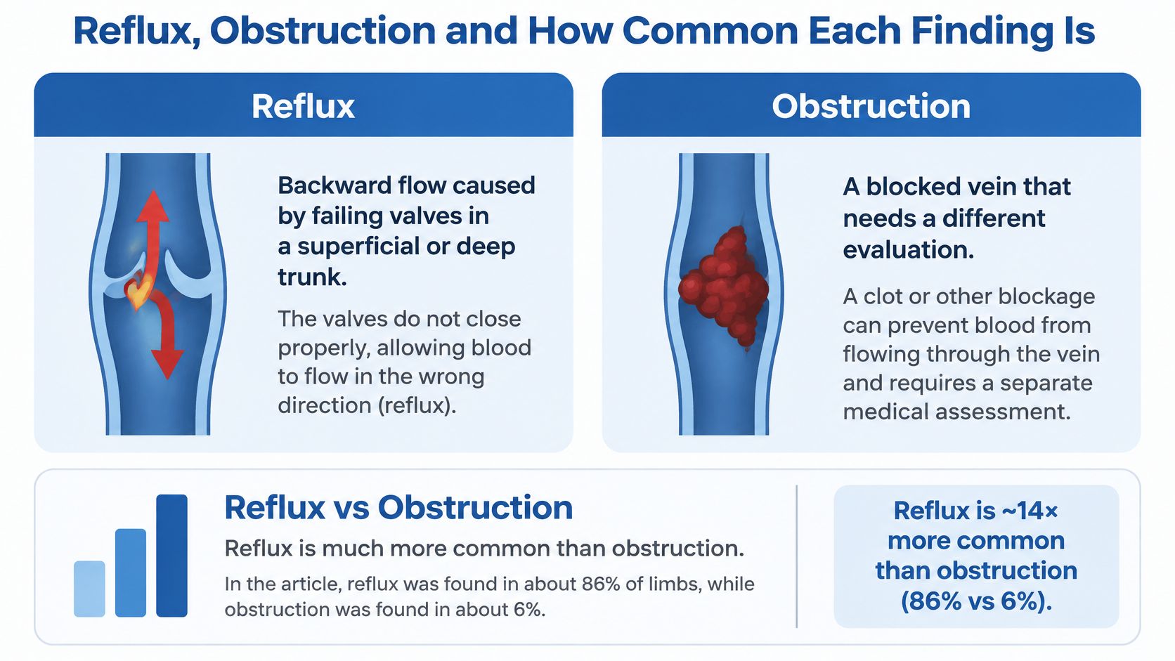
Reflux and obstruction affect blood flow in different ways. Reflux is a direction problem: the vein stays open, but faulty valves let blood fall backward and pool, raising pressure in the lower leg over time. Obstruction means the vein is narrowed or closed, making it harder for blood to pass through. Because the two findings have different effects on drainage, they call for different evaluation and treatment decisions.
| Finding | Population | Reported rate |
|---|---|---|
| Venous reflux in at least one leg | US healthcare workers | 82.1% |
| Venous obstruction | US healthcare workers | 0.2% |
| Superficial venous reflux | Dutch adults, median age 54 (n=2,510) | 23.7% |
| Clinically significant chronic venous disease (CEAP C3-C6) | Dutch adults, median age 54 (n=2,510) | 3.0% |
Why the Rates Don't Match
The last two rows show why an ultrasound finding needs context. In the Dutch population shown, superficial reflux was more common than clinically significant disease. CEAP classes C3 through C6 describe swelling, skin changes such as discoloration or thickening, healed ulcers, and active open ulcers.
A reflux finding does not, by itself, mean advanced disease. Some people have measurable backward flow without swelling, skin damage, or wounds. Others with similar findings have aching or heavy legs and skin changes. The scan describes blood flow; symptoms and a physical examination help show whether that finding matters clinically. The healthcare-worker figure above is best read in that context.
A Reading Order for Your Own Report
When your results arrive, read past the word “reflux” and look at how the findings fit together:
- Reflux, obstruction, or both: Identify which flow problem the scan describes.
- Which segment: Note the named vein and how much of it is involved.
- Does it match your leg: Compare the finding with your symptoms, swelling, and any wound location.
For example, reflux in a vein that serves the area above an ulcer may be relevant to treatment planning. A finding in an unrelated segment, alongside symptoms elsewhere, calls for more questions about the connection. The scan maps the veins; interpreting that map alongside the person's symptoms and examination gives it meaning.
Why Duplex Ultrasound Is the First Test for Venous Disease
A physical exam is like hearing a knock from outside an engine. You know something's wrong, but you can't see which part is failing. Duplex ultrasound is like opening the hood and watching the fluid move. It combines two kinds of information in one examination, and that's the reason it became the first test rather than a backup.
The first half is anatomy. Standard ultrasound imaging shows where each vein runs, how wide it is, and whether its walls compress. The second half is physiology. Doppler measurement shows which direction blood moves, how it responds when the calf is squeezed, and whether valves hold or leak. A purely structural scan gives you only the first half. It can show a wide, twisted vein beautifully and still tell you nothing about whether that vein is the source of your symptoms.
According to the American College of Phlebology imaging guidelines, a complete venous duplex evaluates vein patency, compressibility, diameter, flow direction, obstruction, and valve competency across the deep, superficial, and perforating systems. That same guidance identifies lower-extremity duplex ultrasound as the primary diagnostic modality for chronic venous disease and recommends using it to confirm reflux before treatment.
What Symptoms Can't Tell Your Clinician
Here's the practical problem duplex solves. Leg pain, edema, heaviness, skin discoloration, and ulceration look similar on the surface no matter what's causing them. Superficial reflux can produce that picture. So can deep reflux. So can obstruction, and so can processes that aren't venous at all. Pressing on a swollen ankle and looking at bulging veins can't separate those causes, and each one leads to a different plan.
Your symptoms tell your clinician that venous pressure is too high somewhere. They don't say where the pressure starts.
That's why guidance from NICE and the European Society for Vascular Surgery both place duplex before intervention. NICE recommends confirming the extent of truncal reflux by duplex before treating it, and ESVS names full lower-limb duplex as the primary imaging test when chronic venous disease is suspected or clinically evident. If you're reviewing the basics of the condition itself, our overview of chronic venous insufficiency symptoms, causes and treatment covers how those surface signs develop.
The payoff is a concrete decision. When a clinician can see that one superficial trunk leaks from the groin down while the deep system stays open and competent, treating that trunk makes sense. When the scan instead shows a deep blockage, the conversation shifts completely. At Vein and Wound Institute, a vein consultation and duplex ultrasound help identify where abnormal reflux is occurring and which treatment is most appropriate, because that source, not the visible bulge, drives the plan.
How to Prepare for and Undergo the Scan
A venous ultrasound uses no radiation or injected dye, and it does not require a needle. The sonographer may ask you to change position or help trigger blood flow, because some findings are easier to measure when the veins are filled. Knowing what each step checks can make the exam easier to follow.
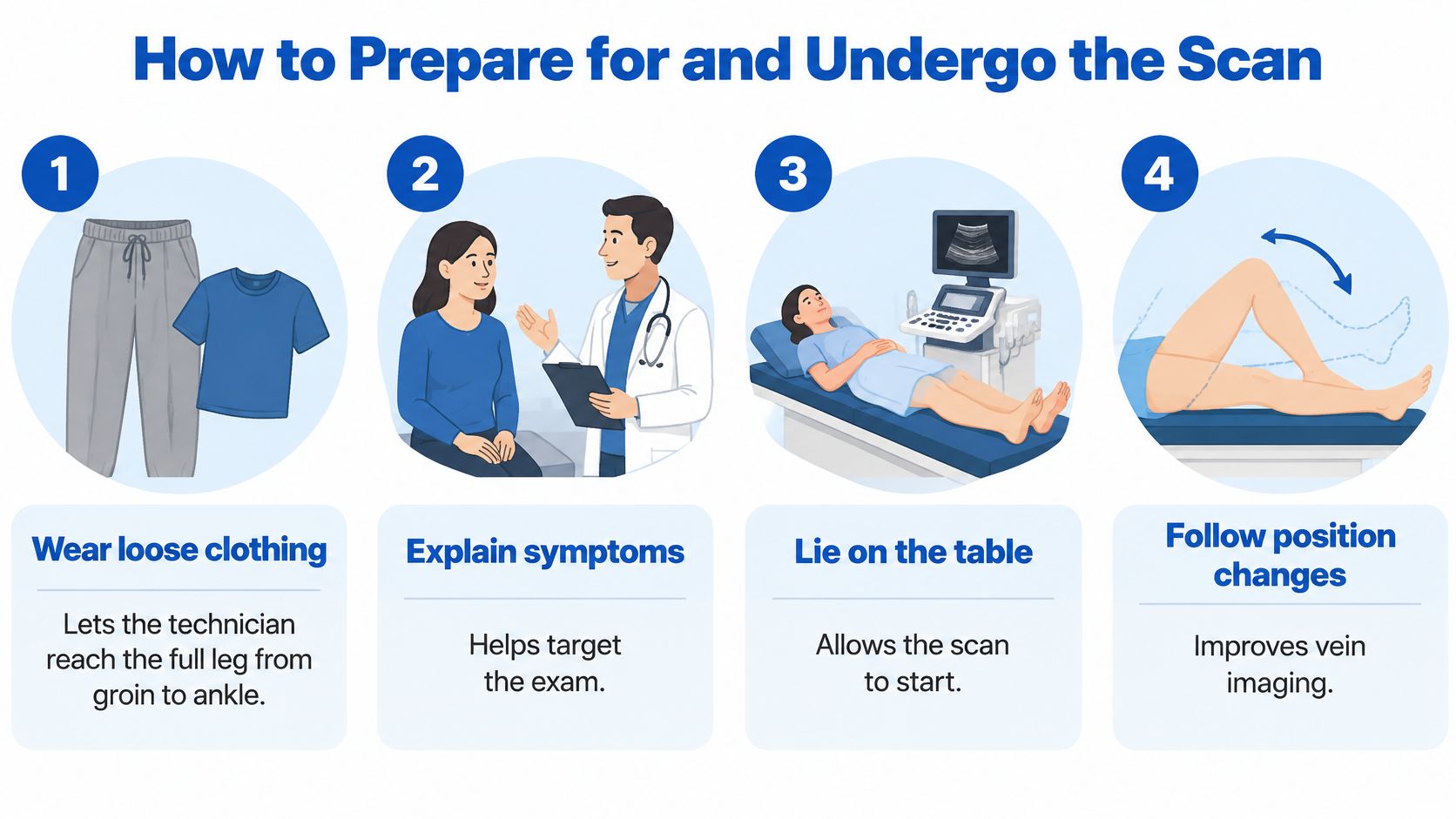
Wear clothing that gives access to your leg. The sonographer may need to scan from your groin to your ankle. Shorts or loose trousers that can be rolled up make this easier.
Share relevant medical history before the scan. Mention prior vein surgery, blood clots, ulcers, and current wounds. This information can help the sonographer decide which areas need particular attention.
Stand if you can do so safely. The scan is often performed upright, with the knee slightly bent, because gravity fills the leg veins. The Society for Vascular Ultrasound lower extremity venous insufficiency protocol recommends this position. If you cannot stand safely, tell the sonographer so they can adapt the exam. Position matters when comparing vein diameters: a 2025 review in the Vascular and Endovascular Review reported mean differences of 19 to 24% in saphenous-trunk diameter between standing and lying positions.
Stay still during the deep-vein checks. The sonographer presses the probe at intervals along your leg to see whether each vein compresses. This helps assess the deep veins and check for thrombus.
The sonographer compresses your calf, then releases it. You may also be asked to cough or bear down. These actions move blood through the veins; the ultrasound records whether it flows backward after the pressure is released.
A proper reflux measurement records the flow sequence. It captures backward flow, the return of forward flow, and any backward flow after release. Brief reversal caused by probe pressure itself does not count as reflux.
Ask what the report will include. You can ask whether it identifies the affected segment, its length, the junction involved, and any obstruction found. These details help explain what the scan shows.
What a Technically Adequate Exam Includes
The study should document vein compressibility, assess for thrombus or obstruction, record Doppler waveforms, measure provoked reflux, and map its source and pathway. Seeing varicose veins on the screen is only part of the exam. The scan needs to show what is happening beneath the visible veins.
Ultrasound findings guide the next step; they rarely settle the diagnosis alone. An adequate exam provides a clear map of the veins it assessed.
What Ultrasound Cannot Tell You
Most explanations of this test end at its strengths. That's a problem, because three common assumptions about venous ultrasound are wrong, and each one can lead to a poor treatment decision.
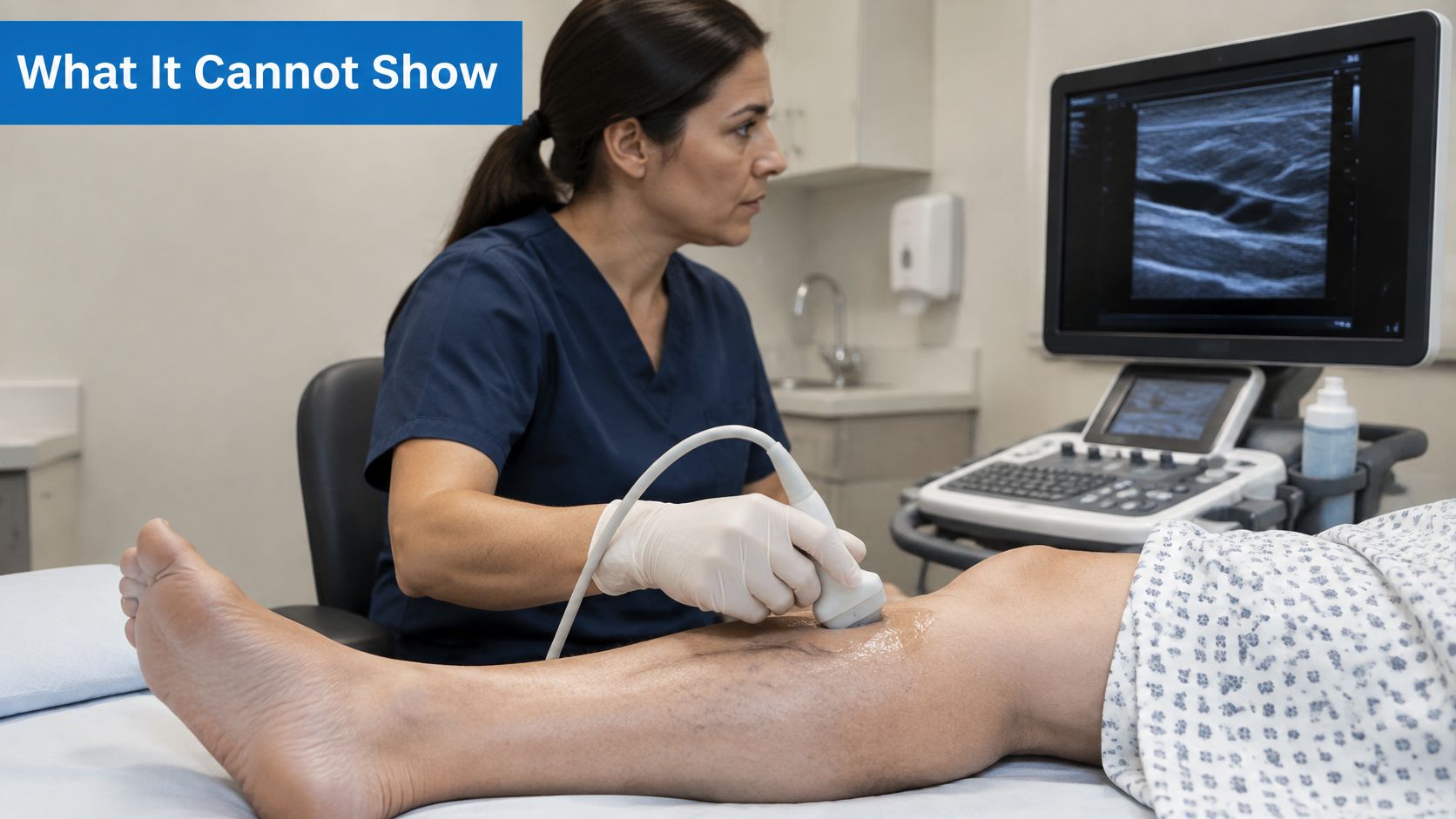
Assumption One That It Checks Your Whole Circulation
A venous duplex characterizes veins. It doesn't establish whether your arteries deliver enough blood to heal a wound or tolerate compression. Those are separate questions with separate tests. Feeling for a foot pulse doesn't bridge the gap either. According to NICE supporting documentation on leg ulcer assessment, pulse palpation alone is unreliable, and ankle-brachial pressure index, toe pressures, or another validated arterial test should be used before compression decisions.
This matters most if you have diabetes, kidney disease, a smoking history, or advanced age, and it's critical with mixed arterial and venous ulcers. Venous reflux and peripheral arterial disease coexist more often than patients expect. Treating the reflux doesn't remove the ischemic risk. Say your duplex shows clear superficial reflux and you have a healing ulcer near your ankle. The reflux finding supports treating that vein. It does not, by itself, clear you for firm compression. You still need the arterial test first. If swelling is part of your picture, our page on chronic venous insufficiency edema explains why the cause of the swelling has to be pinned down before compression is chosen.
Assumption Two That a Normal Leg Scan Rules Everything Out
Duplex is excellent in the leg and limited above it. Pelvic veins and some deep vessels sit where bowel gas, body habitus, and depth degrade the image. As the StatPearls review of venous insufficiency evaluation notes, duplex may not adequately visualize pelvic veins or some deep vessels because of anatomy and technical limitations.
So a normal or limited leg scan doesn't close the case when symptoms outrun the findings. Ask about further imaging, such as cross-sectional studies, if you have:
- Marked one-sided swelling: One leg substantially larger than the other without a leg-level explanation.
- Recurrent ulcers: Wounds that return despite apparently adequate treatment.
- Pelvic or abdominal symptoms: Discomfort suggesting the problem sits higher than the scan reached.
- Prior deep-vein thrombosis: A clot history that raises the chance of proximal obstruction.
Assumption Three That the Test Is Equally Accurate Everywhere
Technique, positioning, and equipment all move the result. A 2025 BMJ Open protocol plans a paired diagnostic study of 84 patients and a randomized trial of 264 patients comparing conventional duplex, assumed to have 60% sensitivity, against ultrasound tomography plus Doppler, expected to reach 80%. Those are protocol assumptions for a planned comparison, not a verdict on your scan. But they underline an honest point. Conventional duplex is the standard first test, and researchers still see room to improve on it. Longer reflux also isn't a severity score, so don't let a big number frighten you or a small one reassure you out of proportion.
From Duplex Map to Treatment, Follow-Up and Next Steps
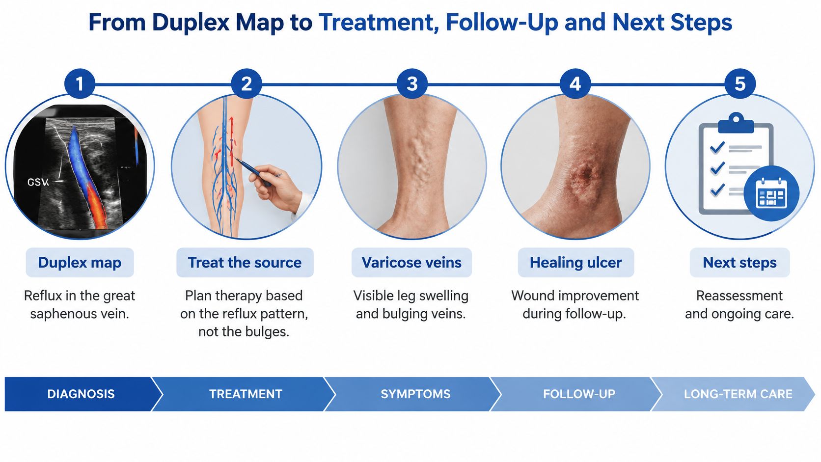
Treatment follows the source of the problem, not the veins you can see bulging at the surface. Follow one typical patient through the process and the logic becomes clear.
Say you arrive with ropey varicose veins down your calf and an ulcer above your inner ankle that's healing slowly. The obvious targets seem to be the bulges and the wound. Your duplex tells a different story. The deep veins compress fully and carry normal flow. The great saphenous vein, the main superficial trunk running up your inner leg, refluxes from the groin junction to below your knee. The calf bulges are tributaries fed by that leaking trunk, and the sustained pressure from the trunk is what's keeping the skin above your ankle inflamed and slow to close.
So the plan targets the trunk, not every bulge. Close the source, and the pressure feeding both the tributaries and the wound bed falls. Treat only the visible bulges, and the leaking trunk repressurizes the leg within months.
The Four Questions the Map Answers
According to NICE guidance on varicose veins, duplex ultrasound is the recommended confirmatory and treatment-planning study for suspected primary or recurrent varicose veins, rather than relying on physical examination alone. The map it produces answers four questions. Does reflux exist? Which truncal vein or junction is incompetent? How far does the reflux extend? Does deep obstruction or thrombus change management?
That fourth answer can redirect everything. Persistent superficial reflux sustains ambulatory venous hypertension, edema, skin inflammation, and ulcer recurrence, which is why closing a leaking trunk helps wounds. Isolated deep obstruction or mixed disease calls for a different strategy and can limit how much weight the superficial findings carry.
How Endovenous Treatment Uses the Map
Modern treatment closes the problem vein from within. Through a small catheter or access point, the clinician seals the refluxing trunk along the segment the duplex identified. Once that vein closes, blood naturally redirects through healthier veins, which reduces venous pressure and improves symptoms. This differs from traditional stripping, which removed the vein surgically through incisions.
The choice among endovenous options depends on the size and location of the vein, your ultrasound findings, your symptoms, and your overall vascular health. If radiofrequency ablation of the saphenous trunk is recommended, our guide to radiofrequency ablation of the greater saphenous vein and recovery walks through what to expect afterward.
Follow-Up in Four Steps
- Baseline duplex: The original map documents the refluxing segment and the state of the deep system before anything is done.
- Treatment: The identified source is closed, and wound care or compression continues in parallel where appropriate.
- Early closure check: A follow-up ultrasound confirms that the treated vein has closed and that flow has rerouted as intended.
- Monitoring if symptoms persist: Ongoing heaviness, swelling, or a stalled wound triggers reassessment of the whole picture, not an automatic repeat procedure.
Persistent symptoms after treatment are a reason to remap and reconsider, including arterial supply and proximal disease. They're not automatically a reason to treat another vein.
Throughout, every ultrasound finding should be correlated with your ulcer location, edema, skin changes, and prior procedures. A refluxing segment that doesn't correspond to your wound deserves a second look before it deserves a procedure. Insurance approval for vein treatment also leans on this documentation, since medical necessity rests on demonstrated reflux and symptoms. If the administrative side is unfamiliar, understanding prior authorization in 2026 explains how that approval step works before a scheduled procedure.
What to Bring When You Request an Evaluation
Arrive with a written symptom list, your wound history including how long any ulcer has been open, records of prior vein procedures, and notes on diabetes or known circulation problems. Then ask for three specific things. A duplex covering the deep, superficial, and perforating veins. A report naming the refluxing segment, its length and junction, and any obstruction. And, if you have an ulcer, an arterial test before compression is selected.
At Vein and Wound Institute, we offer vein consultation, duplex ultrasound, treatment options matched to the map, and follow-up ultrasound to confirm closure, with wound care coordinated alongside vein treatment when both problems are present. Your single next step is simple. Confirm your scan covered the deep veins and perforators, and request a report that lists reflux durations and segments by name. Everything else in your plan builds on that document.
If your legs are heavy, swollen, or carrying a wound that won't close, Vein and Wound Institute can perform a complete duplex ultrasound that maps your deep, superficial, and perforating veins and pairs it with the arterial assessment your compression and wound decisions require. We'll explain your reflux times and segments in plain language, then build treatment around the source the map identifies. Visit Vein and Wound Institute to request your evaluation and bring your symptom list, wound history, and prior procedure records with you.