
Your sock leaves a deep band above the ankle that's still there an hour after you take it off. By late afternoon your calves ache, the skin over your shin looks faintly stained, and one vein running up the inside of your leg has turned thick and rope-like. Compression socks from the pharmacy help, right up until the moment you peel them off.
That pattern, heaviness that builds through the day, swelling that improves overnight, a vein that bulges, is the classic signature of venous reflux in a superficial leg vein. Radiofrequency ablation (RFA) is one of the most common ways to treat it, and in well-selected patients it works very well. It also gets oversold.
The distinction this article keeps returning to is simple. Closing a faulty vein and fixing everything a patient feels are two different outcomes. RFA is measured on the first. Patients live with the second.
Table of Contents
- How Radiofrequency Ablation Closes a Diseased Vein
- Who Qualifies and When Compression Comes First
- How the Procedure Unfolds Step by Step
- How Well RFA Works, Including for Leg Ulcers
- RFA Compared With Surgery and Laser Treatment
- Recovery, Recurrence and Risks to Expect
- Decide Whether RFA Fits and Book a Vein Consultation
How Radiofrequency Ablation Closes a Diseased Vein
The one-way door that stops closing
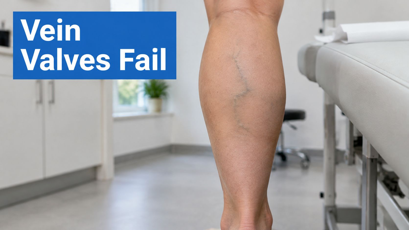
Leg veins carry blood upward against gravity, and they depend on a ladder of one-way valves to do it. Each valve opens as blood rises, then snaps shut behind it so the column cannot fall back. When the leaflets of a valve in a superficial vein stop meeting in the middle, the door hangs open. Blood surges back down, pools, and stretches the vein wall under steady pressure.
That backward flow is the reflux behind the heaviness and swelling. The vessel it distends is the bulging vein you can see, and the one involved most often is the great saphenous vein, which runs from the groin down the inner leg to the ankle.
What the catheter does
Radiofrequency ablation treats that vein from the inside. The skin is numbed with local anesthesia, and a thin catheter goes in through a needle-sized entry point, usually below the knee or near the ankle, then advances up the faulty segment under continuous ultrasound. Once positioned, the catheter delivers radiofrequency energy that heats the vein wall. The collagen in the wall contracts, the lumen collapses, and the treated segment seals shut. Blood reroutes through the healthy deep and connecting veins, and the body gradually absorbs the closed segment over the following months.
Nothing is pulled out. No groin incision, no surgical removal, no general anesthesia. That is why recovery looks so different from traditional vein stripping, which takes the vein out through incisions.
The technique has been refined for more than two decades. Radiofrequency ablation became an important minimally invasive alternative to surgical vein stripping as endovenous technology matured in the late 1990s and early 2000s, and second-generation catheters produced substantially stronger short-term results. A systematic review of endovenous radiofrequency treatment reported ablation rates close to 100% at 6-month follow-up, with no major adverse events in the studies evaluated. Larger registries then tracked treated patients for as long as five years, and durability held: clinical recurrence after RFA stayed well below what followed conventional surgery or laser ablation. Well below is not zero. Residual disease and new reflux in other segments are why ultrasound surveillance after treatment is standard rather than optional.
A sealed vein is also a narrower achievement than it sounds. Closure is the outcome an ultrasound can confirm at follow-up. Whether the aching, the swelling or the skin staining settles is a separate result, one that depends on how much damage the reflux caused before the vein was closed.
One planning principle sits underneath all of this: treatment aims at the source of the reflux, not at the veins visible at the skin's surface. A bulging tributary can be a symptom of a faulty valve 20 centimeters away, which is why the catheter goes where the ultrasound points rather than where the eye looks.
In one sentence: radiofrequency ablation closes the faulty vein from the inside with heat, so blood has to find a healthier route.
Who Qualifies and When Compression Comes First
Not everyone with a visible vein needs a procedure. Plenty of people have mild reflux that never progresses to skin damage, and plenty more get enough relief from support stockings, leg elevation, and regular walking that an office procedure never becomes necessary.
What puts someone on the candidate list
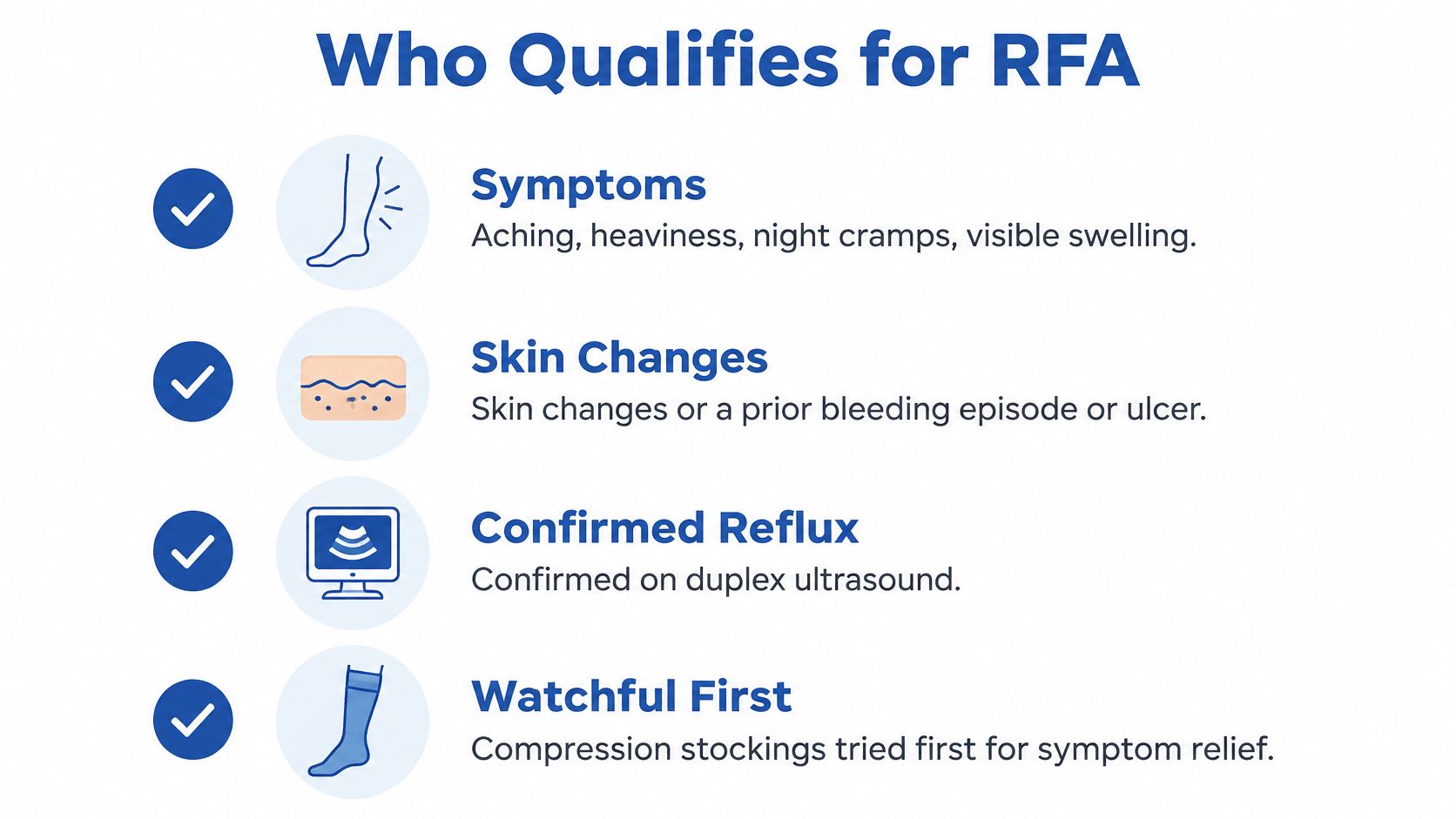
The typical candidate has symptomatic reflux confirmed on duplex ultrasound: aching that worsens through the day, heaviness, night cramps, visible swelling, or skin changes such as brownish staining and eczema-like dryness around the ankle. A history of a vein that bled through the skin, or a healed or active venous ulcer, pushes the decision toward treatment rather than observation.
The ultrasound is what separates a cosmetic complaint from a medical one. It shows which valves fail, where the reflux starts and stops, how the deep veins are working, and whether the vein is large enough and straight enough to take a catheter. That mapping is why a duplex ultrasound evaluation of chronic venous insufficiency comes before any decision about technique.
Several factors narrow the field. Vein size and location determine whether a catheter is even feasible. Deep venous health matters, because closing a superficial vein when the deep system is blocked can make swelling worse. Arterial circulation gets checked before treating anyone with a wound, since poor arterial flow changes the whole plan. Diabetes, mobility, and the presence of infection all shift the risk and benefit calculation.
When stockings are the right first step
For many patients, conservative care comes first, sometimes because symptoms are mild, sometimes because reflux hasn't been demonstrated, and sometimes because an insurer requires a documented trial. Our approach follows the same logic: conservative therapy may be recommended first depending on symptoms, ultrasound findings and insurance requirements, and not every patient needs a procedure.
A minimally invasive procedure becomes reasonable when venous disease causes persistent symptoms, or when compression, elevation and activity modification don't provide enough relief.
A fair trial means weeks, not days. Graded compression worn consistently for four to six weeks tells you something. A pair of socks worn twice and abandoned doesn't.
Two situations usually postpone treatment rather than cancel it. Pregnancy is one, because vein pressures and anatomy change and often settle after delivery. Active skin infection is the other, since nobody should have a catheter placed through inflamed tissue. The physician confirms eligibility in person, with the ultrasound images on the screen.
How the Procedure Unfolds Step by Step
A single vein usually takes under an hour, and most patients walk out and go home the same day. The order below matters because each stage sets up the next, and two of them are where good results are most often lost.
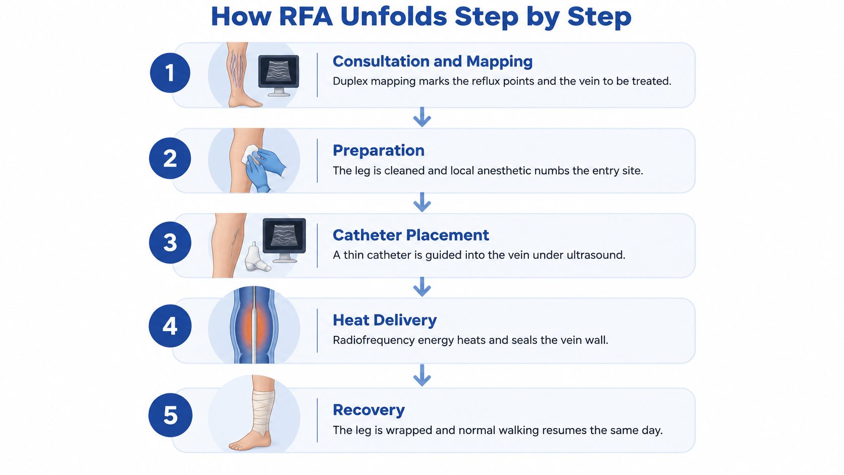
1. Consultation and duplex mapping. The reflux points and the exact vein segment to be treated are marked on the leg before anything is sterilized. Treating only the visible bulge leaves the engine of the problem running, so the map decides what the catheter will close.
2. Local anesthesia at the entry site. The skin where the needle goes in is numbed. Most minimally invasive endovenous procedures are performed under local anesthesia, and patients usually remain awake. Expect pressure or brief discomfort rather than significant pain, and treat any promise of a completely sensation-free procedure with skepticism.
3. Tumescent anesthesia along the vein. A dilute anesthetic solution is injected into the tissue surrounding the vein. It does two jobs: insulating nearby nerves and skin from the heat, and compressing the vein snugly around the catheter. Rushing this step is the most consequential slip in the procedure, because thin insulation is how skin burns and temporary nerve irritation happen.
4. Catheter placement. The catheter enters through a needle-sized opening and is advanced to the top of the faulty segment under continuous ultrasound, so the operator watches the tip the whole way. Many treatments are performed through exactly this kind of needle-sized entry point rather than open surgery. When a segment curves sharply, the ultrasound view has to be re-established before the catheter moves again.
5. Heat delivery during slow withdrawal. The catheter is pulled back at roughly 2 to 3 mm/s while the wall is heated to 85 to 120 °C, a protocol reported in a technical review of endovenous radiofrequency ablation. Ultrasound should show the wall contracting as the catheter moves. Withdraw too fast and a segment escapes treatment, which is how residual reflux survives an otherwise careful procedure.
6. Confirmation and dressing. Duplex imaging confirms the vein has collapsed with no flow inside it, and the entry point gets a dressing. Technical success is defined by those duplex findings rather than by how the leg feels on the table. Published series report a wide spread of occlusion rates in the first months of follow-up, which is why on-table verification carries real weight. Closure confirmed today is the start of surveillance, not the end of it.
7. Compression and walking. A stocking goes on and walking is encouraged soon afterward, since movement keeps blood traveling through the deep veins while the superficial route is closed. The physician sets any limits on strenuous exercise or other activities based on the specific procedure performed.
Questions worth asking before the day
- Which veins will be treated, and is the great saphenous vein one of them or is this a tributary-only plan?
- How many sessions are planned, and what determines whether a second one is needed?
- How will closure be confirmed, and when is the follow-up duplex scheduled?
For symptomatic axial great-saphenous-vein reflux, current vascular guidelines support either thermal or nonthermal ablation, extending treatment from the groin to below-knee segments when clinically indicated and technically appropriate, as the same endovenous ablation technique review sets out. Heat, glue, and foam are technical options chosen after the ultrasound map, and the vein's size, location, and your symptoms drive the choice.
How Well RFA Works, Including for Leg Ulcers
A vein that reads closed on duplex and a leg that feels better are two different endpoints, and the published figures measure the first far more often than the second. Keep that gap in mind as the numbers below come up.
Published RFA outcome benchmarks
| Evidence point | Reported result | Context to keep in mind |
|---|---|---|
| Pooled anatomical success, small saphenous vein | 96.0% (95% CI 91.6% to 98.2%) | Systematic review with moderate heterogeneity across studies |
| Success across treated limbs | Pooled RFA estimate below the single-vein figure, with stripping and foam sclerotherapy close behind | Large multi-study synthesis; definitions of success varied between reports |
| Occlusion in a randomized comparison | Closure maintained through two years of follow-up | Patient satisfaction trailed the closure rate at the same time point |
| Venous leg ulcer, perforator RFA vs compression | Healing or marked improvement favored perforator RFA over compression alone | Post-thrombotic ulcers; recurrence also ran lower with RFA |
| Venous leg ulcer, RFA plus compression vs compression alone | No statistically significant overall healing advantage in a 56-patient randomized trial | Healing trended faster and recurrence lower with RFA |
Why the pooled numbers disagree
Those figures are ranges drawn from groups of patients, and individual results still vary. A systematic review of small saphenous vein treatment reported pooled anatomical success of 96.0% for RFA, with a 95% confidence interval of 91.6% to 98.2% and moderate statistical heterogeneity, meaning results differed between studies and settings rather than clustering around one neat number.
Widen the lens and the estimate moves. When reviewers pool a far broader set of reports, spanning many thousands of treated limbs across well over a hundred studies, the RFA figure lands lower than that single-vein result while stripping and foam sclerotherapy sit close behind. That spread reflects real differences in which vein was treated, how long patients were followed, which catheter generation was used, and how each study chose to define success. A pooled average is an average of definitions as much as an average of outcomes.
The ulcer question
Patients with a venous leg ulcer ask a sharper question than "does it close the vein." They ask whether the wound will heal, and the evidence there is more qualified than a single success percentage suggests.
A randomized comparison of ablation and compression for venous ulcers in 56 patients found no statistically significant difference in overall ulcer healing between compression alone and RFA plus compression, although healing was faster and recurrence was lower with RFA, and two patients developed type 1 endovenous heat-induced thrombosis. The systematic review summarized in the same analysis concluded that endovenous ablation plus compression improves healing compared with compression alone, with less certain evidence on recurrence and complications, particularly for ulcers that have lasted more than six months.
Perforator disease reads more encouraging. In post-thrombotic ulcers, one trial found perforator RFA produced healing or marked improvement more often than compression alone, with recurrence running lower during follow-up.
The accurate verdict: RFA can accelerate healing and reduce recurrence in selected patients when it is integrated with compression and with treatment of the wound itself. Ulcer closure still depends on factors no catheter can reach.
Ulcer duration, deep venous disease, arterial circulation, infection, diabetes, and mobility all move the expected outcome, and a treated vein can reopen or a new segment can reflux over time. That is why an active wound needs a venous leg ulcer assessment alongside the vein plan, followed by surveillance rather than a single visit.
RFA Compared With Surgery and Laser Treatment
A patient with a refluxing great saphenous vein usually hears three names: radiofrequency ablation, surgical stripping, and endovenous laser ablation. All three aim to take the diseased vein out of circulation. The differences that matter in daily life are how the vein is reached, what anesthesia is needed, how long the closure lasts, and how quickly you recover.
RFA, surgical stripping and laser ablation compared
| Factor | Radiofrequency ablation | Surgical stripping | Endovenous laser ablation |
|---|---|---|---|
| Access to the vein | Catheter through a needle-sized entry point | Incisions at the groin and along the leg | Catheter through a needle-sized entry point |
| Typical anesthesia | Local anesthesia with numbing around the vein | Associated with general anesthesia | Local anesthesia |
| Five-year clinical recurrence | 18.7% | 34.6% | 38.6% |
| Typical recovery | Return to normal daily activity within a short period | Longer healing at incision sites | Daily activity within days; bruising along the treated vein is common |
| Scarring | Uncommon at very small access points | Visible incision scars possible | Uncommon at very small access points |
What the comparison does and doesn't tell you
The access row is the one patients feel first. RFA and laser both work from inside the vein through a puncture, while stripping removes the vein through open incisions at the groin and down the leg. That single difference drives the anesthesia row too: office-based endovenous work avoids the larger incisions and general anesthesia associated with traditional vein stripping, so most patients stay awake under local numbing and feel pressure or brief discomfort rather than significant pain.
The recurrence row deserves the most caution. The three percentages come from a single five-year randomized comparison of the techniques, and they describe groups of treated patients, so read them as an ordering rather than a forecast for your leg. In that comparison, RFA recorded the lowest five-year recurrence of the three options. The figures also count recurrence as it is defined clinically: reflux detected on ultrasound, symptoms that return, and veins that sometimes need no further procedure at all. Two patients behind the same number can have very different follow-up stories. Figures like these exist because closure is verified by ultrasound over years, and follow-up scans continue whichever technique you choose.
Laser ablation works on the same principle as RFA with a different energy source, and it carries its own after-effects worth understanding before you commit. Anyone weighing the two should read about endovenous laser ablation side effects so the comparison rests on real recovery experiences.
A practical way to use this table: bring it to a consultation and ask which row your provider expects to apply to your legs, and why. The choice depends on the size and location of the vein, what the ultrasound shows, your symptoms and your overall vascular health. If the answer is vague, ask to see the ultrasound images and have the reflux points pointed out.
Recovery, Recurrence and Risks to Expect
The first two weeks
Expect bruising and tenderness along the treated segment, sometimes a firm cord under the skin where the vein is scarring down, and swelling that settles with elevation and walking. The cord can feel alarming the first time you touch it. It is the treated vein doing exactly what it was heated to do, and it typically softens over the following weeks. Access points are needle-sized or a few millimeters wide, so significant scarring is uncommon.
How fast you get back depends on what your day demands. Someone at a desk often resumes work quickly, while a job built on lifting or long hours on your feet may need clearance from the physician first. Strenuous exercise may be limited for a short window while the vein seals. Walking is encouraged, because calf muscle contractions keep blood moving through the deep system while the superficial vein closes.
The line between expected and concerning is worth memorizing.
Call the clinic for: rising calf pain or warmth, sudden swelling of the whole leg, new skin changes, fever, or bleeding at the entry point that doesn't stop with pressure.
The risk list is short but real: heat-related thrombosis in a nearby vein, temporary nerve irritation close to the treated segment, skin burns, pain, and residual reflux if a segment didn't close. Each sits in the minority of cases, and temporary effects like nerve irritation and bruising tend to fade over time, but they belong in the consent conversation before the procedure.
When closure doesn't hold
Closure on ultrasound means the procedure did its mechanical job. Whether your symptoms resolve, and stay resolved, is a separate question that depends on the rest of the circulation. In five-year follow-up data, recurrence was usually found at scheduled scans rather than reported by the patient: 61% of recurrent great saphenous veins and 79% of recurrent small saphenous veins were identified within six months of regular follow-up, and incompetent perforating veins were reported as an important recurrence site needing reoperation, according to a review of RFA outcomes and surveillance.
That is the argument for keeping duplex appointments even when the leg feels fine. It also explains why swelling sometimes persists after a technically clean closure: untreated tributaries, deep venous reflux, lymphedema, obesity, and immobility all keep producing fluid regardless of what happened to one superficial vein.
For anyone whose main complaint is swelling or a slow-healing wound rather than a bulging vein, this distinction matters most. RFA removes one driver of venous hypertension. Compression, exercise, weight management, and follow-up address the rest, and they usually stay relevant after a successful procedure.
Decide Whether RFA Fits and Book a Vein Consultation
You can move this from reading to deciding in three steps.
1. Record your symptoms for several weeks. Note when the heaviness peaks, whether swelling improves overnight, any skin staining or itching around the ankle, and what compression has or hasn't done. A short written log is more useful to a clinician than a memory of "my legs hurt."
2. List the conservative care you've already tried, with dates. Physicians and insurers both ask, and a documented trial of graded compression carries more weight than a vague recollection. If you haven't given stockings a fair run, that's the honest starting point.
3. Book a consultation with duplex ultrasound. Venous evaluation and duplex imaging are what identify where abnormal reflux is occurring and which treatment is most appropriate, whether that turns out to be radiofrequency ablation, another endovenous method, or continued conservative care.
Before you book, run the screen. Symptoms lasting months rather than days. Reflux documented on imaging. A genuine trial of compression behind you. Healthy arterial flow, especially with a wound. No active skin infection. A realistic commitment to follow-up, including the duplex check that confirms closure.
Bring the questions from the procedure section, and add one more: what happens if symptoms persist after treatment? The answer tells you whether the plan accounts for the tributaries, perforators, and deep system that a single catheter can't fix.
Patients with an active wound may also need a wound-care assessment in the same breath as the vein plan, since healing depends on both. Treatment plans are personalized, and radiofrequency ablation is a medical procedure whose fit has to be confirmed by a clinician who has examined your legs and seen your ultrasound.
Vein and Wound Institute treats venous disease and the wounds it causes as one problem, so the reflux behind a heavy, swollen leg and the ulcer that won't close get evaluated together rather than in separate silos. If you're weighing radiofrequency ablation, the process starts with a consultation and duplex ultrasound to map where the reflux begins, and continues with duplex follow-up to confirm the treated vein has closed. Visit Vein and Wound Institute to schedule an evaluation at a location convenient to you in Los Angeles, Santa Barbara, or Ventura County.