
You notice a yellow patch on your leg while drying off after a shower. You look closer, compare it with the other leg, and wonder whether it's a fading bruise, a skin problem, or something happening beneath the surface. Before reaching for a cream, check what surrounds it: swelling, warmth, pain, a raised bump, or an open sore can tell you more than the shade itself.
Table of Contents
- What Yellow Spots on a Leg Really Mean
- The Four Biological Sources of Yellow Discoloration
- Reading What Your Skin Is Telling You
- When Circulation Is the Hidden Cause
- Red Flags That Mean Call Your Doctor Today
- How Doctors Work Up the Cause
- Treatment Paths and What to Expect
- Quick Takeaways and Common Questions
What Yellow Spots on a Leg Really Mean
Yellow spots on a leg describe an appearance, not a diagnosis. Several substances can give skin a yellow tone, including lipids, elastin, and bilirubin. A dermatology review of yellow skin disorders explains why similar colors can arise through different biological processes.
Think of the color as a stain on a ceiling. The stain tells you something has changed, but it doesn't tell you whether the source is a leaking pipe, condensation, or an old repair. Skin color works the same way: you need context before choosing a solution.
Start with the surroundings
A useful first inspection goes beyond asking whether the patch is lemon-yellow, orange, or yellow-brown. Notice its shape, texture, location, and what your leg feels like.
- Surface: Is the area flat, raised, firm, crusted, or open?
- Distribution: Is there one mark, a cluster, or discoloration across a broader area?
- Nearby changes: Do you see ankle swelling, prominent veins, redness, or damaged skin?
- Other symptoms: Is there itching, pain, unusual warmth, fatigue, or yellowing of your eyes?
These observations don't establish a diagnosis. They help you describe the problem accurately and decide whether it needs urgent attention or a scheduled examination.
Similar colors can mean different problems
For example, a flat yellowing area after a known injury raises a different question from several firm yellow bumps without an injury. A discolored ankle with swelling also deserves a different assessment from widespread yellowing that includes the eyes.
You don't need to identify the pigment yourself. Your job is to notice the pattern and avoid assuming that every yellow mark is either harmless or dangerous.
Practical rule: Ask “What else is happening?” before asking “Which cream removes this?”
That shift is especially important when a patch doesn't hurt. Absence of pain doesn't establish that a lesion is cosmetic, particularly when it's persistent, raised, enlarging, or accompanied by other changes.
The Four Biological Sources of Yellow Discoloration
Yellow-looking areas can arise from deposits within the skin, substances circulating in the blood, pigment left after blood-cell breakdown, or material on a wound's surface. Those mechanisms aren't interchangeable.
Deposits and circulating pigment
Lipid deposits can form xanthomas, yellow-to-orange lesions containing macrophages filled with fats. Macrophages are cells that collect and process material; when lipid-laden cells accumulate, they can create a visible bump or plaque. The connection with cholesterol or triglyceride disorders is important, but a bump's appearance alone can't establish an abnormal blood result.
Bilirubin belongs to a different pathway. Your body produces it when red blood cells break down. Think of this as a recycling system: when production, processing, or drainage becomes disrupted, bilirubin can accumulate in the blood and cause jaundice.
Unlike an isolated deposit, bilirubin-driven yellowing generally affects broader areas and may involve the whites of the eyes. It can follow a face-to-body pattern rather than starting as one leg spot, as described in this clinical overview of jaundice and its causes.
The supplied illustration below concerns tooth discoloration, not leg skin. Its dental categories shouldn't be used to interpret a leg lesion.
Tissue staining and wound material
The remaining categories involve iron-containing staining and yellow material on the surface.
- Hemosiderin: This pigment contains iron left after red blood cells break down in tissue. It can produce rust-colored, brown, or yellow-brown areas. An analogy is a rust stain beneath an old leak: the visible mark remains where material escaped.
- Wound exudate or crust: Yellow material in or around an open lesion may be wound fluid or crust rather than pigment. Warmth, pain, drainage, spreading redness, and delayed healing change its significance.
A healing bruise is another possible explanation for a localized yellow patch. It shouldn't become the default explanation when there's no clear injury history or when the area is changing unexpectedly.
Location and timing narrow the question. A persistent ankle stain, a newly raised nodule, and rapidly developing widespread yellowing call for different investigations, even if you would describe all of them as yellow.
Reading What Your Skin Is Telling You
The most useful sorting question is often simple: Is this a stain, a deposit, a plaque, or wound material? You can describe those features without trying to diagnose yourself.
A stain changes color without creating an obvious raised structure. A bump projects above the surrounding skin. A plaque is a broader area of altered skin, while a crust sits on its surface, sometimes over a wound.
Appearance-based triage of yellow leg spots
| Appearance | Likely Source | What to Notice | Typical Next Step |
|---|---|---|---|
| Flat yellow-brown stain | Possible blood-breakdown pigment or venous staining | Ankle location, swelling, itching, visible veins | Clinical examination, with circulation assessment when indicated |
| Raised yellow bumps or yellow-red-brown plaques | Possible lipid deposits or an inflammatory skin condition | Firmness, multiple lesions, enlargement, front-of-shin location | Medical or dermatologic assessment; selected blood tests or biopsy |
| Yellow crust within an open lesion | Wound exudate, crust, or possible infection | Warmth, drainage, pain, redness, delayed healing | Wound assessment; urgent care when red flags accompany it |
| Widespread yellowing | Possible bilirubin accumulation | Yellow eyes, dark urine, pale stools, abdominal or systemic symptoms | Timely medical evaluation focused on jaundice |
The “likely source” column lists possibilities, not conclusions. A photograph or mirror inspection can help you communicate the appearance, but it can't reliably distinguish every condition in the table.
Plaques aren't all cholesterol deposits
Necrobiosis lipoidica can produce yellow-red-brown plaques on the front of the legs and is strongly associated with diabetes. Diagnosis is often clinical, although uncertain appearances may require biopsy, as discussed in this clinical discussion of a leg rash.
That association doesn't mean every shin plaque proves diabetes. It means the clinician should consider the skin findings together with your medical history rather than treating the color in isolation.
For a concrete comparison, a firm yellow bump with intact skin raises questions about a deposit. A similarly colored area that is wet, tender, and open raises questions about a wound. They may look alike from a distance, yet the next steps differ substantially.
Don't scrape a crust to decide what's underneath or squeeze a raised lesion to test it. Describe what you can see and let the examination resolve uncertainty. If several appearances overlap, such as staining around an open ankle sore, tell the clinician about both rather than focusing only on the yellow portion.
When Circulation Is the Hidden Cause
A yellow-brown ankle patch accompanied by swelling deserves attention as a possible circulation problem. The lower “gaiter” region, the area around the ankle and lower calf, is a characteristic location for venous skin changes.
In chronic venous insufficiency, valve reflux or obstruction can raise pressure in the leg veins. That pressure reaches small blood vessels, allowing red blood cells to escape into surrounding tissue. After those cells degrade, iron-containing pigment remains, producing the staining described in venous leg assessment and management guidance.
How a vein problem reaches the skin
Think of vein valves as one-way elevator doors. When they function properly, they help prevent backward movement. When they don't close effectively, blood can flow backward and pressure can build below.
The analogy has a limit: the pigment isn't blood sitting on the skin's surface. It develops within tissue after cells have escaped and broken down. That's why a circulation-related stain can't be approached like something you wash away.
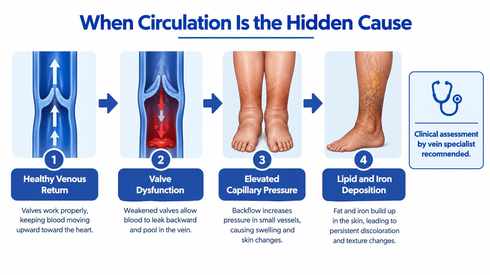
The venous-staining mechanism illustrated here centers on iron deposition. Lipid deposits are a separate explanation for yellow lesions, not a required stage of venous disease.
The symptom cluster matters
Venous staining becomes more clinically meaningful when it occurs with other lower-leg changes:
- Fluid buildup: Ankle or lower-leg edema accompanies the discoloration.
- Inflamed skin: Itching or stasis dermatitis affects the surrounding area.
- Visible vein changes: Varicose veins appear near the affected region.
- Skin breakdown: A wound develops, particularly around the inner ankle.
A clinician assesses the whole leg, including swelling, skin integrity, temperature, and pulses. This overview of chronic venous insufficiency symptoms, causes, and treatment provides further context for a circulation-focused visit.
Arterial circulation requires separate consideration. Cool skin or diminished pulses raises concern about blood supply reaching the tissues, rather than blood returning through the veins. Both problems can coexist.
Compression isn't a color treatment. Its safety depends on the circulation beneath the skin.
That distinction prevents a common mistake: buying strong compression because a discolored ankle looks venous, without first checking whether arterial blood flow is adequate.
Red Flags That Mean Call Your Doctor Today
Urgency comes from accompanying symptoms, not yellow color alone. A stable, painless raised bump generally belongs in a different category from a painful wound with spreading redness.
Use the checklist below to decide what kind of help to seek. If symptoms are severe or you feel acutely unwell, seek urgent medical care rather than waiting for a routine appointment.
Seek same-day assessment
- Spreading redness or fever: These can accompany infection, especially around an open lesion.
- Severe pain or black tissue: A wound with these findings needs urgent evaluation for tissue damage and its cause.
- Pain with cool skin: A discolored area with coolness or suspected poor blood flow needs prompt assessment.
- Yellow eyes or rapid widespread yellowing: These warrant medical review, particularly with dark urine, abdominal pain, fever, or confusion.
- An open lesion that isn't healing: Arrange assessment rather than repeatedly trying different creams.
For additional context on blood-supply concerns, see peripheral arterial disease and poor circulation.
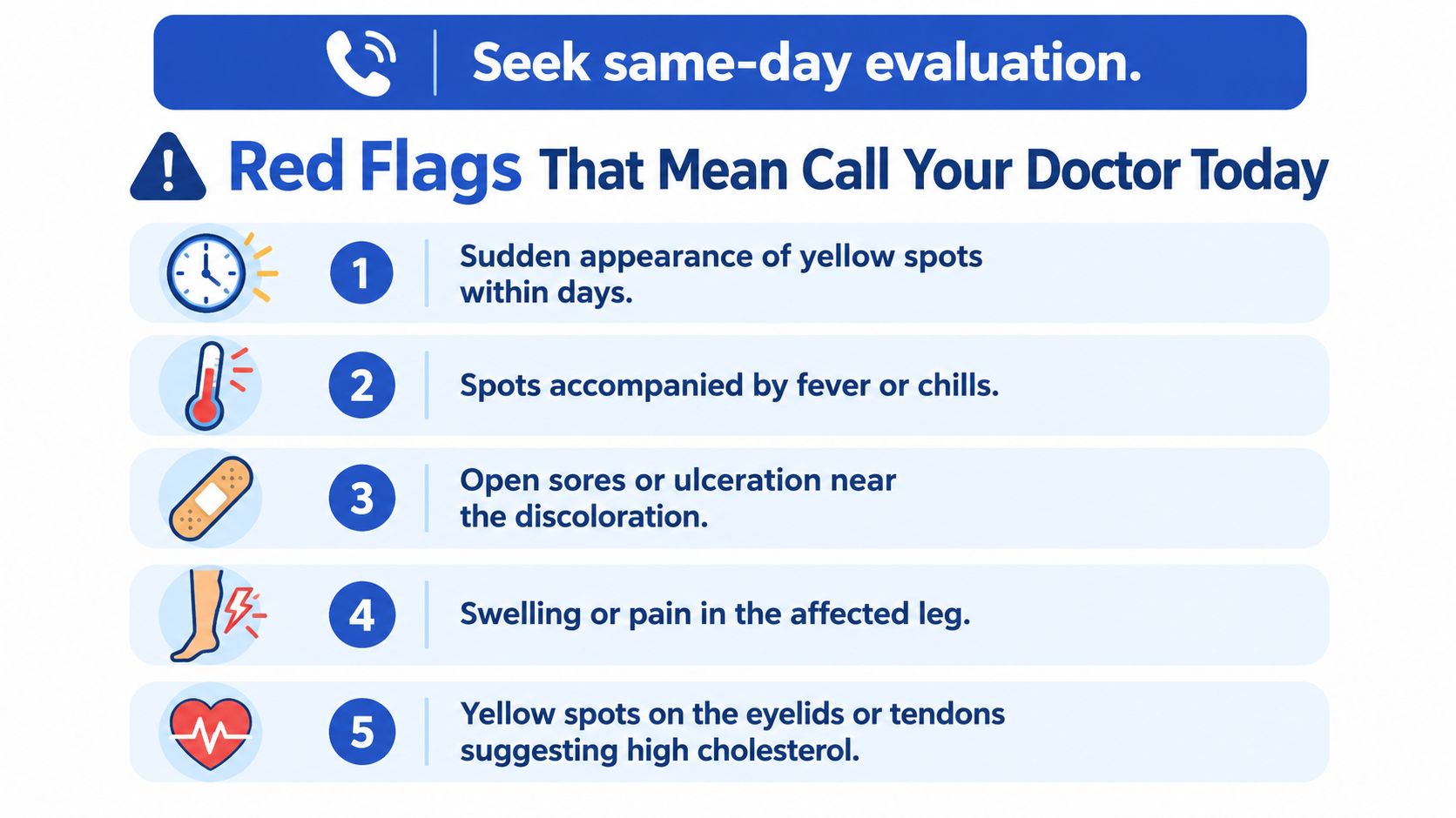
Not every finding in the illustration independently requires same-day care. Stable yellow bumps without acute symptoms usually warrant a prompt scheduled evaluation, while fever, severe pain, rapid deterioration, or concerning wound changes increase urgency.
Schedule a visit soon
Persistent, enlarging, recurrent, raised, or multiple lesions deserve examination even when they don't hurt. Discoloration with ankle swelling, heaviness, itching, or prominent veins also calls for a review of the surrounding skin and circulation.
You don't have to wait until a wound appears before asking for help. Equally, you don't need to assume the worst because a patch is yellow.
Save this checklist:
Urgent today: Severe pain, fever, spreading redness, black tissue, cool painful skin, or yellow eyes with concerning symptoms.
Book soon: Persistent or changing lesions, raised bumps, multiple spots, or discoloration with swelling.
Bring: A description of when it began, how it changed, and your current medication list.
How Doctors Work Up the Cause
A useful visit starts with an examination, not an automatic order for every possible test. The clinician looks at distribution, texture, warmth, swelling, pulses, and any open areas, then chooses investigations that answer a specific question.

Different appearances lead to different investigations
| Clinical Question | Possible Evaluation | What It Helps Clarify |
|---|---|---|
| Could a raised lesion be a xanthoma? | Lipid testing and medical or family history | Whether abnormal lipid metabolism may be involved |
| Could a shin plaque be diabetes-associated? | Review of diabetes history and assessment guided by the examination | Whether the skin finding fits a broader metabolic picture |
| Could widespread yellowing be jaundice? | Bilirubin and liver-function testing | Whether bilirubin accumulation or liver-related disease needs investigation |
| Could ankle staining reflect venous disease? | Duplex ultrasound | Whether reflux or venous obstruction is present |
A lipid panel measures blood fats; it doesn't identify every yellow lesion by itself. Likewise, liver tests are selected because the overall pattern suggests jaundice, not because one patch looks yellow.
Checking whether compression is safe
The ankle-brachial index, or ABI, compares blood-pressure measurements at the ankle and arm. Think of familiar inflatable blood-pressure cuffs being used to assess whether the leg's arterial supply needs closer investigation.
Published guidance generally supports standard or high compression at an ABI of approximately 0.8–1.3 when significant arterial disease is absent; 0.5–0.8 usually calls for supervised modified compression, while below 0.5 generally contraindicates routine high compression, according to venous-ulcer guidance on compression and perfusion.
Some conditions can make ABI falsely high, so clinicians may use toe-pressure testing instead. The result must be interpreted alongside the examination, not as a stand-alone permission slip.
For a visit prompted by swelling, this explanation of whether leg swelling can indicate a vein disorder can help you prepare. Bring your medication list, relevant family history, and observations about the lesion's progression; biopsy is reserved for situations where tissue examination would clarify uncertainty.
Treatment Paths and What to Expect
Treatment should address the cause, not merely cover the color. Repainting a stained ceiling doesn't repair a leaking pipe, and removing a visible skin lesion doesn't necessarily resolve an underlying circulation or metabolic problem.
A diagnosis also helps establish a sensible goal. For one person, the priority may be closing a wound; for another, it may be investigating lipid abnormalities or preventing further skin damage.
When the problem is venous or wound-related
For confirmed venous disease, the plan depends on the circulation findings and whether skin breakdown is present. Compression may form part of care when arterial assessment supports it. A specialist may also consider minimally invasive treatment of the affected veins rather than focusing solely on the pigment.
Available vein-treatment categories include endovenous laser ablation, radiofrequency ablation, and ultrasound-guided foam sclerotherapy. The choice requires examination and imaging; a yellow patch alone doesn't establish that a procedure is appropriate.
An open lesion needs a wound-specific plan. Depending on its cause and condition, care may involve:
- Wound-bed management: Clinician-directed debridement when indicated.
- Dressings and protection: Selection based on the wound rather than its color alone.
- Infection management: Assessment and treatment when infection is suspected.
- Follow-up: Monitoring whether the tissue is making meaningful healing progress.
Specialist reassessment is important when a presumed venous ulcer fails to progress after about six weeks of guideline-conforming care, as noted in the earlier compression guidance. Uncertainty about the wound's cause can also justify reassessment and possible biopsy.
When the source isn't venous
With suspected xanthomas, medical evaluation of lipid metabolism takes priority over cosmetic removal alone. The point is to investigate why deposits formed, not erase the visible evidence.
For a diabetes-associated plaque, the clinician needs to confirm the skin diagnosis and consider the broader medical picture. A treatment plan shouldn't be inferred from a photograph or replaced with an over-the-counter cream.
Jaundice requires treatment directed at its underlying cause. Causes range from benign inherited bilirubin-processing differences to liver injury or bile-duct obstruction, so generalized yellowing isn't a reason to start a leg-skin treatment.
Ask what success will look like for your diagnosis. Symptom relief, improved wound condition, and management of the underlying disorder may matter more than rapid disappearance of discoloration.
Quick Takeaways and Common Questions
Read the shape, location, and symptoms together. Keep a short record of changes so you can give the clinician more useful information than “it looks yellow.”
Not all orange-brown leg marks are venous stains. Pigmented purpuric dermatoses can produce orange-red-brown flat spots and pinpoint purpura, while venous hypertension can cause pinpoint bleeding over diffuse staining, as described in this review of pigmented purpuric dermatoses. Similar appearances are a reason for examination, not a reason to guess.
Common questions
Can yellow spots be the first visible sign of diabetes or abnormal cholesterol?
Yes, some associated lesions can prompt investigation before someone knows about an underlying disorder. They don't prove either diagnosis, and blood testing should follow the clinical assessment.
Why can compression be the wrong first move?
A leg can have both venous and arterial disease. The safety assessment described earlier determines whether substantial compression is suitable and what level a clinician should recommend.
How long does venous staining take to fade?
There isn't a dependable timetable you can infer from color alone. Don't treat fading as the only measure of success; ask your clinician how they'll assess circulation, symptoms, and skin health.
When is a biopsy worth considering?
Biopsy can help when the appearance is uncertain or a lesion or wound doesn't behave as expected. It isn't automatically necessary for every yellow patch.
Your next step should match the problem you can observe: urgent care for red flags, a scheduled examination for persistent lesions, and circulation or wound assessment when swelling or skin breakdown accompanies discoloration.
For adults whose leg discoloration comes with swelling, visible veins, or a non-healing sore, Vein and Wound Institute offers clinical examination, onsite circulation diagnostics, vein treatment, and full wound care. Visit the practice to arrange an evaluation and discuss a plan that addresses the underlying cause as well as the skin changes.