
You finish a long day, sit down, and your calves feel stretched from the inside. Maybe it's just tired muscle. Maybe your skin itself has changed. Learning to tell those two apart is the first step toward knowing what to do next.
Table of Contents
- When Tight Skin on the Legs Is More Than a Sore Muscle
- How Circulation and Lymphatics Shape the Feel of Your Skin
- The Five Conditions That Cause Tight Skin on Legs
- A Guided Self-Check You Can Do at Home
- What a Vein and Wound Specialist Looks For Next
- Treatment Options That Match the Cause
- When Tight Skin on the Legs Warrants a Same-Week Visit
- What to Take Away About Tight Skin on the Legs
When Tight Skin on the Legs Is More Than a Sore Muscle
Someone finishes a long shift on their feet, peels off their socks, and finds calves that feel stretched and stiff. By morning, the feeling's gone. That's a familiar, mechanical kind of tightness, and it often follows hard exercise, prolonged standing, or a day when you drank too little water.
Contrast that with a different patient. She notices the same stretched feeling every evening, not just after busy days. Her shins look shinier than they used to, one ankle stays puffy, and the skin near it feels firm when she presses it. Nothing hurts sharply, so she hesitates to call it a medical problem.
The Pattern Matters More Than a Single Evening
Tight skin on legs can be temporary and mechanical, or it can be the surface sign of a circulatory, lymphatic, or skin problem underneath. Muscle soreness usually tracks with activity and fades. Skin that's changing tends to repeat, progress, or settle into one leg more than the other.
Three questions sort most cases quickly:
- Is this new or recurring: A one-off feeling after a workout differs from a pattern you can predict each evening.
- Is it one-sided: One leg behaving differently from the other deserves more attention than both legs feeling tired together.
- Is the skin itself changing: Shine, firmness, color change, or thickening points away from plain muscle fatigue.
If only the muscle aches, rest usually answers the question. If the skin's texture, color, or shape is changing, rest won't explain it.
That distinction guides everything that follows. Everyday tightness needs reassurance and simple habits. Persistent tightness with visible skin change needs a name for its cause, because the right treatment depends entirely on which system underneath is struggling.
How Circulation and Lymphatics Shape the Feel of Your Skin
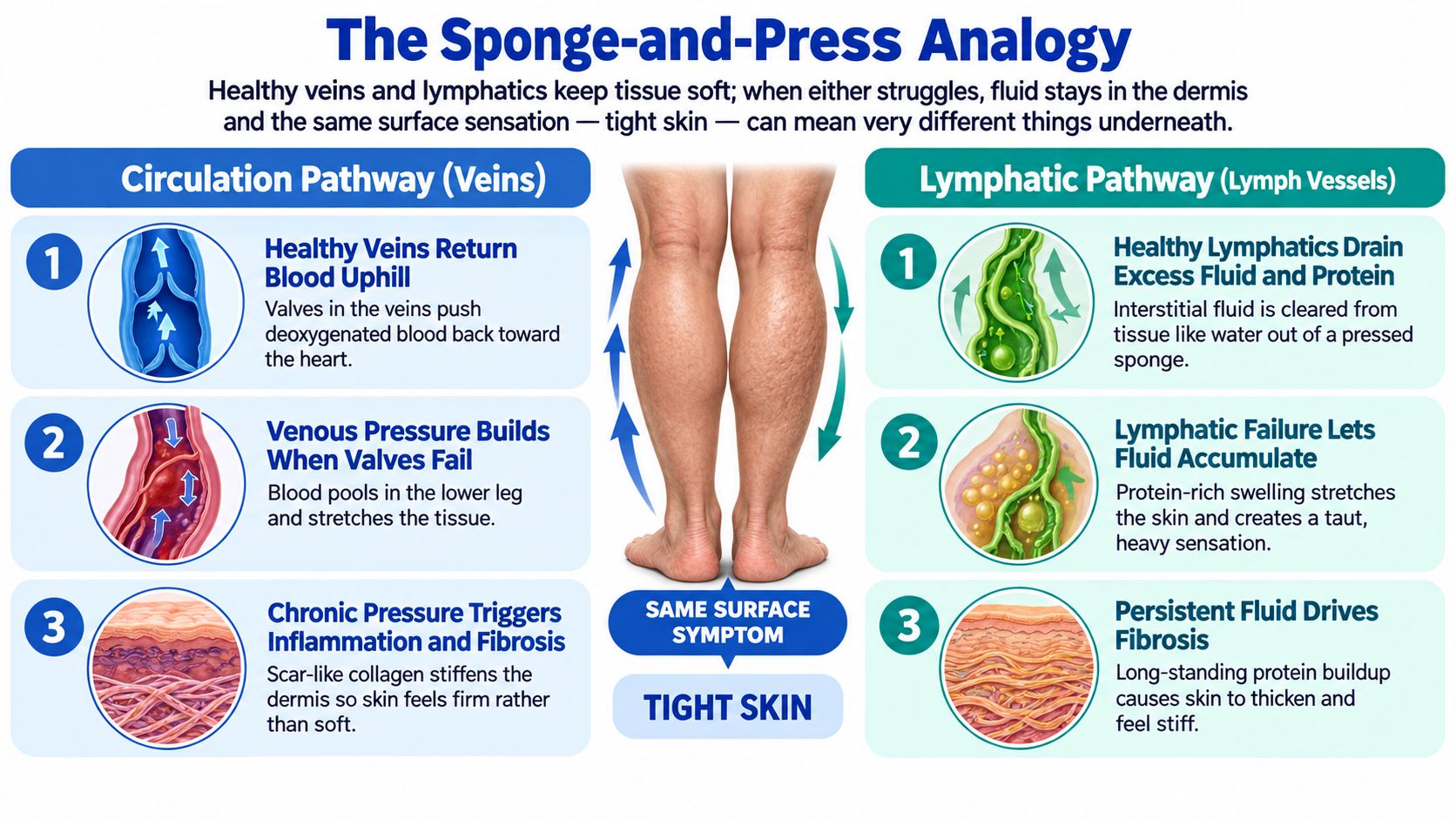
Think of the tissue under your skin as a sponge. Healthy veins return blood uphill toward the heart, and healthy lymphatics drain excess fluid and protein out of that sponge. When either system struggles, fluid stays trapped, the sponge saturates, and your skin feels taut, heavy, or stiff.
Three Terms Worth Knowing
Venous pressure is the force inside your leg veins. When vein valves fail, blood pools downward and pressure rises in the lower leg. That pressure pushes fluid into surrounding tissue.
Interstitial fluid is the fluid sitting between cells. A little is normal. Too much stretches the skin from within, which is why swollen legs often look smooth and shiny.
Fibrosis is what happens when fluid lingers. Persistent fluid triggers inflammation, and over time the body lays down scar-like collagen. Skin that was soft becomes firm, and that's a different problem from simple swelling.
Why the Same Feeling Has Different Causes
Tight skin can come from pressure, fluid, scar tissue, or a mix of all three. Two people with similar looking legs may need very different care. One needs fluid moved out. The other needs the failing vein treated before fibrosis advances further.
This is also why clinicians check arterial flow before assuming swelling is venous. Resources on ABI testing for asymptomatic adults explain how ankle pressure measurements screen for arterial disease. For the lymphatic side, the overview of lymphedema evaluation and care describes how drainage failure changes tissue over time.
Hold onto the sponge image. Every condition ahead is a different way that sponge gets overloaded, damaged, or stiffened.
The Five Conditions That Cause Tight Skin on Legs
Each condition below tells a different story inside the leg. Prevalence data show these aren't rare curiosities. In the United States, approximately 3% to 4% of the population has chronic venous insufficiency, according to clinical data on venous insufficiency prevalence. Skin pigmentation changes affect roughly 3% to 13% of people in population studies cited there.
Venous, Lymphatic, and Fat-Related Causes
Chronic venous insufficiency starts when vein valves leak and pressure builds. Over years, that pressure drives inflammation and lipodermatosclerosis, a woody thickening around the ankle, often with brown staining. A detailed explanation appears in this guide to chronic venous insufficiency symptoms and treatment.
Chronic lymphedema begins with protein-rich fluid the lymphatics can't clear. Early swelling pits when pressed. Later, fibrosis makes tissue firm and leathery, pitting fades, and skin can thicken or crack, as described in research on lymphatic fibrosis mechanisms.
Lipedema involves painful, symmetric fatty tissue, usually in both legs, that can feel firm and tender. It typically spares the feet, bruises easily, and doesn't respond to elevation the way fluid swelling does.
Arterial and Autoimmune Causes
Peripheral arterial disease produces tight, shiny skin through the opposite problem, too little incoming blood. Skin becomes thin and atrophic, leg hair thins, and the leg may feel cool. Systemic sclerosis tightens skin through autoimmune collagen buildup. Skin looks shiny and loses elasticity, sometimes involving the toes, with color changes in cold weather.
| Condition | Typical Pattern | Pitting? | Skin Signs |
|---|---|---|---|
| Venous insufficiency | One or both, ankle heavy | Early yes | Brown staining, woody firmness |
| Lymphedema | Often one leg, includes foot | Fades over time | Thick, leathery texture |
| Lipedema | Both legs, spares feet | Minimal | Tender, nodular fat |
| Arterial disease | One or both, distal | No | Thin, shiny, hairless, cool |
| Systemic sclerosis | Symmetric, may involve toes | No | Tight, shiny, less elastic |
A Guided Self-Check You Can Do at Home
This check takes about five minutes and gives you precise observations to bring to an appointment. It doesn't diagnose anything. Its job is to replace vague worry with describable facts.
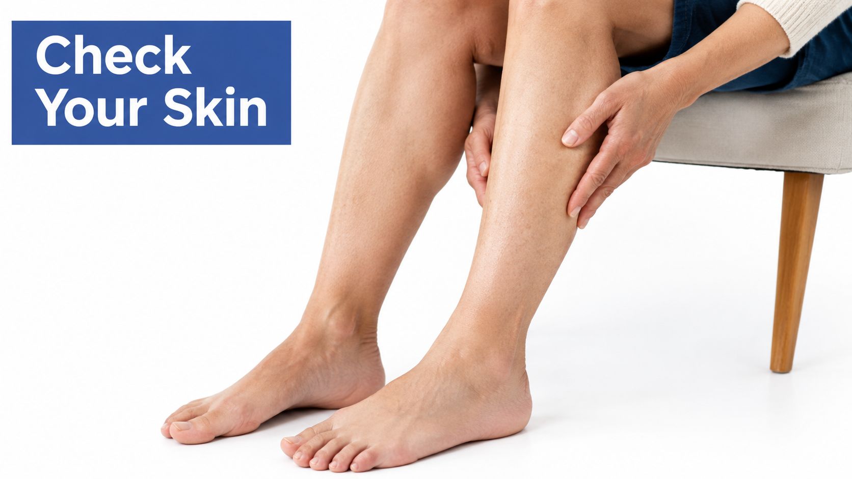
Press, Compare, and Observe
- Test for pitting: Press your thumb into the shin and ankle for five seconds. If a dent remains, note how long it takes to fill back in.
- Compare both legs: Stand in front of a mirror and check size, color, and shape side by side. Asymmetry is information.
- Feel temperature and pulse: Use the back of your hand on each foot. A noticeably cooler foot is worth reporting, as is a pulse you can't find on one side.
- Inspect the surface: Look for hair loss, a shiny sheen, brown staining near the ankle, flaking, or small open areas that won't close.
Then prop your legs on pillows for thirty minutes. If tightness eases considerably, fluid is likely part of the picture. If firmness stays exactly the same, fibrosis or another tissue change may be involved. Guidance on whether leg swelling signals a vein disorder helps interpret that response.
Write it down: Note when it started, what makes it worse, whether elevation helps, and what the skin actually looks like. Those four details save real time in the exam room.
You're not trying to name the condition yourself. You're learning to describe it in sentences a clinician can act on.
What a Vein and Wound Specialist Looks For Next
The clinic visit builds directly on your self-check. The specialist examines skin texture, color, pulses, and temperature, then chooses tests that separate venous, arterial, and lymphatic causes from one another.
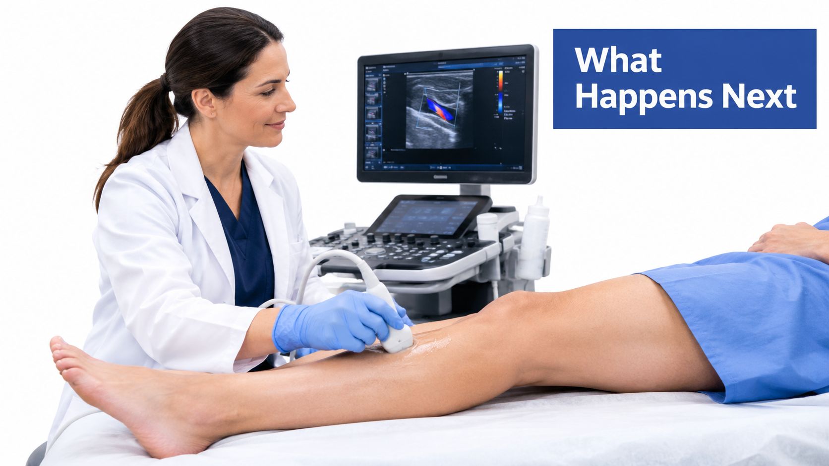
The Tests and What Each One Answers
Duplex ultrasound is usually first. It maps blood flow in real time, showing whether vein valves leak backward, called reflux, or whether a vein is blocked. It covers both superficial and deep veins without radiation or needles.
The ankle-brachial index compares blood pressure at the ankle and arm to screen for arterial insufficiency. A toe-brachial index helps when ankle readings are unreliable. When lymphatic failure is suspected, lymphoscintigraphy or bioimpedance can confirm how well lymph fluid moves.
Epidemiological data underline why this sorting matters. In an analysis of 12,320 individuals, clinically overt chronic venous insufficiency was present in 40.8% of participants, reported in European Heart Journal data on venous disease burden. With numbers that large, guessing from appearance alone misleads often.
Why Arteries Get Checked Before Compression
Compression squeezes the leg to move fluid upward. On a leg with poor arterial inflow, that squeeze can further reduce blood delivery. Documenting arterial status first protects the tissue you're trying to heal.
A board-certified, RPVI-credentialed physician interprets the duplex images personally and reads skin and wound findings alongside them. That integration matters, because treating reflux while managing an active wound in one plan usually beats treating each in isolation.
Treatment Options That Match the Cause
There's no single treatment for tight skin, because there's no single cause. Matching the intervention to the mechanism is the whole point of the workup you've just read about.
Pathways by Diagnosis
For venous fibrosis, treatment targets the leaking vein. Endovenous ablation or VenaSeal closes the faulty segment so pressure drops. Graduated compression, calf muscle activation through walking, and diligent moisturization then support the skin while induration gradually softens.
For lymphedema, care centers on complete decongestive therapy. That combines fitted compression garments, manual lymphatic drainage techniques, skin care, and prompt treatment of any cellulitis. Consistency matters more than intensity.
Lipedema care relies on conservative measures first, including compression and movement, with surgical consultation considered for selected patients. Weight strategies alone don't remove lipedema tissue, which frustrates many patients who've been told otherwise.
Arterial disease requires vascular evaluation before anything else. Tight, shiny skin can indicate chronic peripheral arterial disease with dermal atrophy, and clinicians should document pulses and temperature and obtain an ankle-brachial index before applying compression, as set out in clinical guidance on peripheral arterial disease assessment.
| Cause | Primary Treatment | Realistic Timeline |
|---|---|---|
| Venous reflux | Ablation or VenaSeal, compression | Edema eases in weeks |
| Lymphedema | Decongestive therapy, garments | Ongoing maintenance |
| Lipedema | Compression, surgical consult | Gradual, individualized |
| Arterial disease | Vascular evaluation first | Depends on perfusion |
| Established ulcer | Debridement, grafts, venous care | Months of coordinated care |
Set expectations clearly. Edema often improves within weeks. Fibrosis softens over months. Established ulcers need coordinated debridement, possible grafts, and ongoing venous care to stay closed. Integrated practices that treat the underlying vein problem and the active wound together can shorten recovery and reduce recurrence, because the pressure driving the wound gets fixed while the wound itself is dressed and monitored.
When Tight Skin on the Legs Warrants a Same-Week Visit
Not every tight leg is an emergency, and this isn't a scare list. It's a triage guide. The goal is matching your situation to the right speed of care.
Urgent, Same-Week, and Routine
Seek urgent vascular evaluation for rest pain in the toes, black discoloration, or a foot that suddenly turns pale and cold. Add sudden one-sided swelling with pain and tightness together, or skin that's hot and red, since those combinations can signal clot or infection.
Book a same-week specialist visit for new asymmetry between legs, a wound that hasn't closed in two weeks, brown staining spreading up the shin, or firmness that's clearly progressing. Calf pain that appears reliably with walking and eases with rest also belongs here.
Routine evaluation fits stable, long-standing heaviness without skin change. It still deserves an appointment, just not an alarmed one. You'll get a fuller workup when nobody's rushing.
A few situations lower the threshold further:
- Any skin break in a leg with known arterial disease: Small wounds in poorly perfused tissue can deteriorate quickly.
- Recurrent redness in a swollen leg: Repeated cellulitis suggests lymphatic involvement that needs a prevention plan.
- Tightness with numbness or weakness: Nerve symptoms widen the list of possible causes beyond veins.
If the skin itself is changing in color, texture, or integrity, the leg needs to be seen. That single rule covers most decisions you'll face tonight.
When in doubt between same-week and routine, choose same-week. An unnecessary prompt visit costs an afternoon. A delayed necessary one can cost tissue.
What to Take Away About Tight Skin on the Legs
Three ideas carry the whole subject. If you remember nothing else, remember these.
First, tight skin is a clue, not a diagnosis. The same surface sensation can come from fluid, fibrosis, poor arterial inflow, or autoimmune skin change. Cleveland Clinic's overview of lymphedema progression and skin changes shows how one cause alone can move from soft pitting swelling to firm, thickened tissue, which is why labels matter before treatment starts.
Second, pattern beats severity. A mildly uncomfortable leg that's one-sided, discolored, and progressively firm tells a clinician more than a very uncomfortable leg after a hard workout. Where it is, how long it lasts, and whether the skin looks different outweigh how dramatic it feels.
Third, assessment beats guessing. The right treatment depends on the right cause, and compression on the wrong leg can set healing back. Guessing feels faster. It usually isn't.
If your tightness is persistent, your skin's appearance is changing, or a wound isn't healing, schedule a vein and wound evaluation rather than continuing to self-treat. Bring your self-check notes. Mention which leg, how elevation responded, and when you first noticed the change.
There's a hopeful note underneath all of this. Integrated venous and wound care can address the underlying reflux and its skin consequences together, in one coordinated plan. Legs that feel tight today aren't necessarily legs you're stuck with. They're legs asking a question, and the question has testable answers.
If persistent tight skin, swelling, or a slow-healing wound is part of your daily life, Vein and Wound Institute offers duplex ultrasound evaluation, minimally invasive vein treatment, and advanced wound care across Southern California in one coordinated plan. Visit the site to request an appointment and get a clear name for what's happening in your legs.