
22% of 2,160 people with arm lymphedema reported at least one cellulitis episode in their lifetime, and 11% had one in the previous year, according to a large international study of lymphedema and infection. Cellulitis is a bacterial infection of the skin and the soft tissue beneath it. On a swollen limb it doesn't need a dramatic entry point. A fissure between the toes, a patch of dermatitis, or a scrape the size of a fingernail will do.
That fact reframes the whole subject of lymphedema wound care. A wound on a swollen leg or arm is not primarily a skin problem with some swelling around it. It's a fluid problem with a hole in it, and the hole won't close reliably while the fluid stays.
So every decision here starts from three checks: how well the limb is moving fluid, whether arterial blood is arriving, and how likely infection is right now. From there, the steps are ordered so a patient can carry them out at home, and so a clinician can see quickly when home care has reached its limit.
Table of Contents
- Why Swollen Limbs Heal Wounds More Slowly
- Check the Limb Before Choosing Compression
- Infection Risk and When to Seek Urgent Care
- Compression Options and What Each One Does
- Daily Wound Care Under Compression
- Monitoring Limb Volume and Healing Over Time
- Managing Lymphedema Wounds With Limited Specialist Access
Why Swollen Limbs Heal Wounds More Slowly
A retired teacher clips her shin on the corner of a coffee table. On an ordinary leg, the scratch scabs over in three days. On her swollen left leg, it weeps clear fluid for a week, the skin around it turns shiny and tight, and by day ten the whole calf is warm and red. Nothing about the injury changed. The tissue it landed in did.
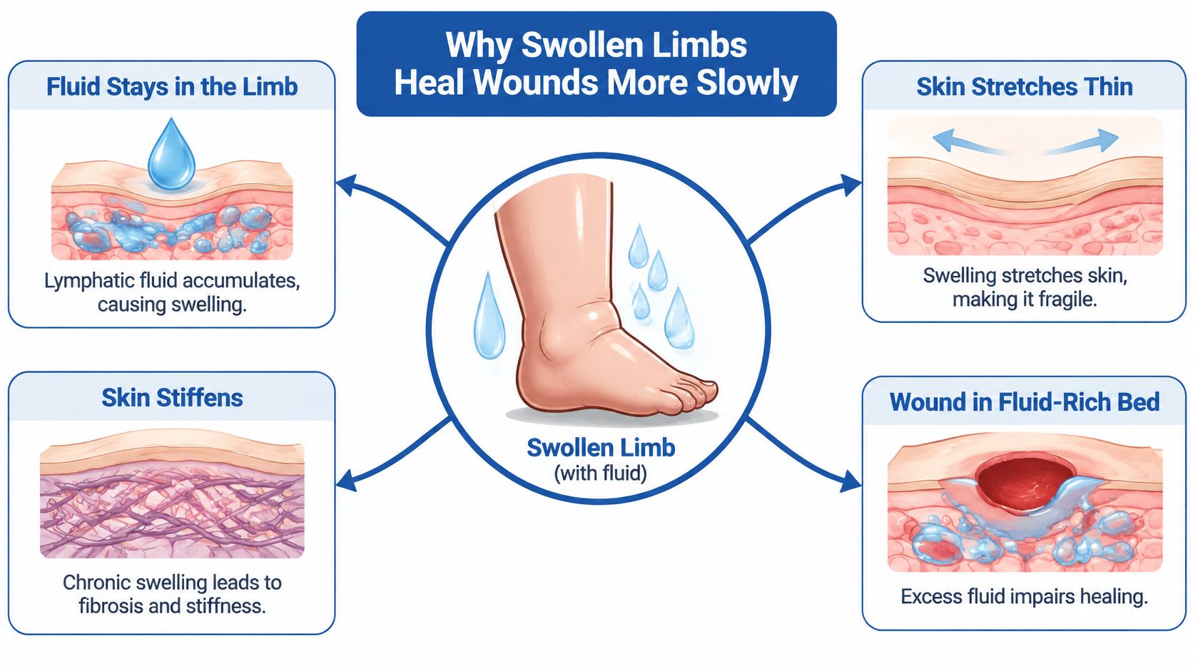
Lymphatic vessels collect fluid from between the cells and carry it back toward the bloodstream. When that return slows, protein-rich fluid stays in the limb. It widens the distance between the smallest blood vessels and the skin cells that need oxygen, so oxygen and nutrients take longer to reach the wound edge. Immune cells that normally patrol that space and clear bacteria move through stagnant, protein-heavy tissue far less efficiently. Skin stretched by fluid thins, dries and splits.
What Fibrosis Does to a Healing Wound
Long-standing swelling is not just extra water. It sets off persistent inflammation, and that inflammation lays down collagen until the tissue stiffens. A fibrotic limb feels firm and woody. Oxygen and nutrients diffuse through it more slowly, and the limb loses its shape, which matters later when a garment has to fit and hold.
The dam comparison. A crack in a dam matters most when the river behind it is high. Patch the crack and leave the pressure, and it opens again in the same place.
The numbers support that comparison. In a large international study of lymphedema and infection, people with well-controlled arm lymphedema had a 46% lower likelihood of recent cellulitis than those with poorly controlled disease, with an odds ratio of 0.54. Risk also climbed with time in that study: compared with lymphedema present for less than a year, the odds of recent cellulitis rose to 2.15 at one to two years, 2.86 at two to five years, and 3.15 at five to ten years.
A wound plan that only changes dressings is aimed at the crack. Swelling control, skin protection, hygiene, rapid treatment of suspected infection and prevention of the next breakdown lower the pressure behind it. Limb volume, skin integrity, drainage, pain and infection history belong in the record alongside wound measurements.
Check the Limb Before Choosing Compression
Compression can reduce swelling, support venous return and help a wound heal, but it has to match the limb in front of you. Consider a cast: the same material can help one injury and harm another if circulation, skin tolerance or infection is ignored. Before a wrap or garment goes onto a swollen limb with an open wound, four questions need answers: arterial supply, infection status, heart status, and skin condition. The table below turns that into a working checklist.
| Finding | What It Suggests | Action Before Compression |
|---|---|---|
| Weak or absent pedal pulses | Possible arterial insufficiency | Vascular assessment before any high compression |
| Active cellulitis or spreading redness | Acute infection | Treat the infection first and hold compression until it clears |
| Decompensated heart failure | Fluid overload risk | Specialist input before any compression |
| Open ulcer with dark, dry or gangrenous tissue | Possible arterial or deep tissue problem | Urgent in-person assessment |
| Stiff, fibrotic skin in a stage II or III limb | Structure needs firm containment | Inelastic or custom system after volume reduction |
| Venous ulcer with reflux | Venous pressure driving the swelling | Compression combined with treatment of the venous source |
| Diabetes with reduced sensation | Higher pressure injury risk | Check skin and garment fit at every change |
Read the top half as safety checks. A weak or absent pedal pulse means the limb may not tolerate high pressure, so arterial supply gets measured before anything is wrapped. Spreading redness means infection may already be moving through tissue. Black, dry or gangrenous tissue in an ulcer needs urgent in-person assessment, not a fresh dressing and another day at home.
The bottom half is about matching the tool to the limb. A stiff, fibrotic stage II or III limb often needs the firm containment of an inelastic or custom system once volume is down. A venous ulcer with reflux needs compression along with treatment of the venous source, because pressure from above keeps pushing fluid and inflammation back into the skin. Diabetes with reduced sensation calls for skin and garment fit checks at every change, since a pressure mark that a fully sensing foot would report can pass unnoticed.
Why More Compression Is Not Automatically Better
The right pressure, format, application technique and monitoring schedule depend on arterial circulation, tissue tolerance, limb shape, fibrosis, exudate, and whether the patient can physically get the device on. That is a clinical judgment, not a product choice. The evidence base is thinner than many patients assume. A review of the occupational therapy evidence on phlebolymphedema identifies a persistent lack of practice guidelines and clinical recommendations specifically for compression in phlebolymphedema, and notes that evidence supporting complete decongestive therapy also remains limited.
That gap is why conservative care comes first. Compression, skin care, exercise, manual lymphatic drainage and pneumatic compression are usually tried before surgery is discussed. In clinical practice, surgery may be considered after a sustained conservative trial has not provided enough control. When swelling travels with venous disease, the picture is chronic venous insufficiency with lymphedema, and both parts need treating. An evaluation can help determine the severity of chronic venous disease. The plan is based on the source of the problem, not on the veins visible at the skin's surface.
Bring five questions to the first visit. Are the pulses palpable? What is the ankle pressure or ankle-brachial index? Has there been an infection in the past year? Is the heart compensating well enough to shift fluid? What did the skin look like at the last assessment? Those answers set the safe starting point before compression is chosen.
Infection Risk and When to Seek Urgent Care
Start with the assumption that a swollen limb gives infection more room to gain ground. An analysis of infection burden in chronic lower-leg edema reports that among 7,477 patients with chronic lower-leg edema, 2,802 people, or 37.47%, had experienced cellulitis at some point in their lives, 15.78% had it within the previous 12 months, 31.2% of those recent cases were hospitalized, and controlled swelling carried an odds ratio of 0.59 for infection. That last point is useful: swelling control acts like lowering the water level around a leaky wall. The limb may still need wound care, but the surroundings become less supportive of repeated problems.
The same source puts the cellulitis rate at 25.4 episodes per 100 person-years in lymphedema, 47.4 per 100 person-years in phlebolymphedema, 8.9 per 100 person-years in breast-cancer-related lymphedema, and 48.0 per 100 person-years in patients with venous leg ulcers and lymphedema. Read those figures as risk categories, not as a personal forecast. They show why an open venous ulcer plus lymphedema deserves closer attention than swelling alone, and why the care plan should keep fluid balance, arterial status, and infection risk in view from the start.
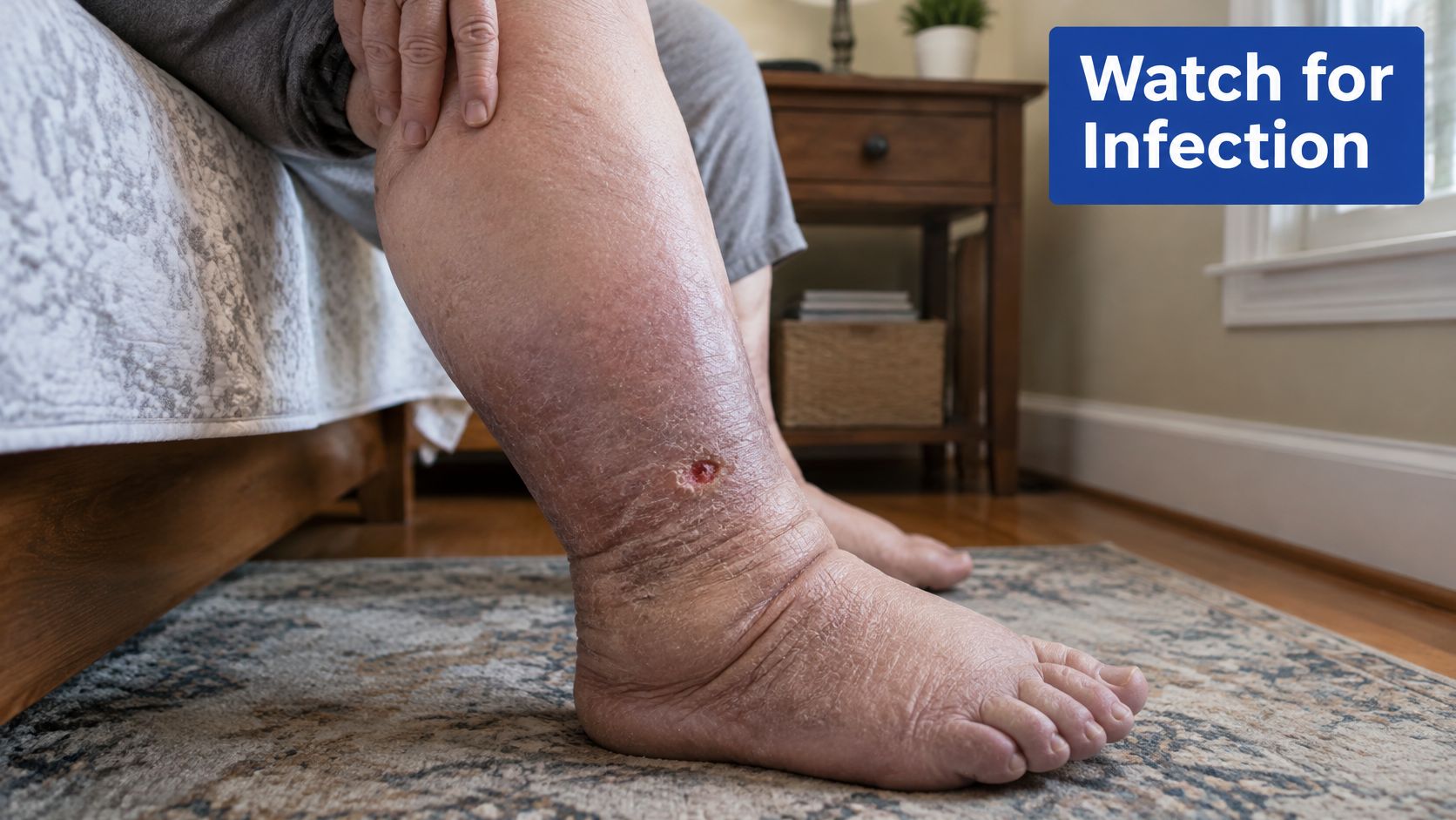
Signs That Need Same-Day or Emergency Care
- Redness spreading faster than a pen mark can be tracked for an hour before it means anything. Same-day assessment.
- Fever, chills, or feeling generally unwell alongside a red limb means the infection may be systemic. Emergency care if there's confusion, rapid heartbeat or low blood pressure.
- Severe pain out of proportion to the wound can signal deep infection rather than surface inflammation. Emergency care.
- Foul odour or cloudy, discoloured drainage points to bacterial load that needs treatment, not a stronger dressing. Same-day assessment.
- A sudden change in limb colour or temperature, hot and dusky or cold and pale, needs urgent in-person review. Emergency care if the limb goes cold, mottled or numb.
- A wound that stops improving over two weeks isn't an emergency, but it does mean the plan needs review rather than another week of the same routine. Same-day assessment.
A red, warm, painful limb on a Saturday is a reason to call, not a reason to wait for a Monday appointment. Early treatment of suspected infection is what helps prevent an admission. The visual checks on signs of wound infection and when to seek medical attention can make that call easier.
Compression Options and What Each One Does
Compression formats aren't interchangeable. Each one delivers a different pressure profile, and each one fails in a predictable way.
| Option | Best Suited For | Main Limitation |
|---|---|---|
| Short-stretch bandages | Reducing limb volume in active treatment | Needs trained application and frequent checks |
| Multilayer bandaging | Complete decongestive therapy in larger volume limbs | Labor-intensive and reapplied daily |
| Inelastic or adjustable wrap | Fibrotic limbs needing firm containment | Needs skilled fitting |
| Custom flat-knit garment | Maintenance after volume reduction | Costly and needs accurate measurement |
| Ready-made compression stocking | Mild swelling and maintenance | Often gives too little containment for stage II or III limbs |
| Pneumatic compression device | Adjunct to bandaging or garments | Cannot replace daily skin care |
How the Two Phases Fit Together
Treatment runs in two phases, and mixing them up is the most common source of confusion. The intensive phase uses multilayer or short-stretch bandaging to bring limb volume down. Lower-stretch systems work here because layering creates a stiff wall that resists the limb as muscles pump, and that stiffness, not raw tightness, is what moves fluid.
The maintenance phase starts once volume is stable. A fitted garment holds the reduction for as long as it's worn, and maintenance compression is lifelong because swelling commonly returns when treatment stops. A device that was right at the start of the intensive phase will often be wrong three weeks later, once the limb is smaller and its shape has changed.
Poor measurement breaks more plans than poor product choice. A garment sized from a stale measurement slips, rolls at the knee, bunches behind the ankle, or leaves a deep mark across the shin. Refitting isn't a sign that the first attempt failed. It's part of the schedule.
Three rules cover most of it. Choose a format the patient or a carer can apply without help, because a perfect device that stays in a drawer does nothing. Check the skin at every change, since pressure injury under compression can start unnoticed. And expect the device to change as the limb changes.
Ask before you buy. Which format suits this limb shape, who will teach the application, how tight should it feel at the toes, how long is it worn each day, and when is the next fitting review?
After a vein procedure, compression stockings may be recommended, and the provider tells the patient the appropriate compression level and how long to wear them. That instruction belongs with the discharge plan, not in a general leaflet.
Daily Wound Care Under Compression
Lymphedema wound care treats the wound and the fluid pressure beneath it at the same time. Complete decongestive therapy combines skin hygiene, manual lymph drainage, multilayer compression bandaging, exercise, and long-term compression garments, as set out in the consensus document of the International Society of Lymphology. The aim is simple: reduce interstitial fluid, improve lymph return through the muscle pump, and limit tissue stiffness that slows oxygen and nutrient movement. Daily care works best when each step protects the skin from both sides. The wound needs a clean, protected surface. The limb needs steady fluid control without new pressure points.
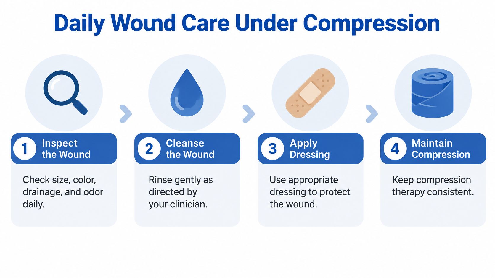
The Eight Steps in Order
- Inspect the wound and the whole limb. Note size, colour, drainage, odour and the state of the skin between the toes and in the folds. Fluid-filled tissue can hide early damage, so look closely at skin that is warm, shiny, cracked, or newly tender. A change you spot on day two is a phone call; the same change on day ten is often an admission.
- Cleanse gently, rinse rather than scrub. Fragile, swollen tissue tears under friction, and every tear is a new entry point for bacteria. Follow your clinician's directions on cleanser and frequency. Let water run over the surface instead of rubbing at slough or crust.
- Dry carefully, particularly between the toes and in skin folds. Trapped moisture softens skin, and softened skin breaks down without any injury at all. Pat rather than drag a towel across the area. Give folds enough air time before dressings or compression go back on.
- Moisturise the intact skin on the limb, never the wound bed. Dry, scaly skin cracks, and cracks let bacteria in. A plain emollient is enough; keep it off the open area. Think of the skin barrier like the seam of a waterproof coat: the fabric can repel water, but a split seam lets moisture and dirt through.
- Protect the skin around the wound with a barrier product. Adhesive dressings lifted off thin, fragile skin take a layer with them. A barrier film or ointment on the surrounding skin keeps removal gentle. This matters when frequent Dressing changes are part of the plan.
- Match the dressing to the drainage. Heavy fluid needs a highly absorbent dressing. A dry wound bed needs one that holds moisture in. The wrong choice leaves maceration around the edges or a stuck dressing that damages the periwound skin. A dressing should manage fluid without turning the surrounding skin soggy or dry.
- Reapply compression as directed, checking it lies smooth. Wrinkles and bunched fabric create pressure points. Make sure the wrap or garment isn't tight at the toes or digging behind the heel. Getting a garment on straight and smooth is a skill worth practising; our guide to how to put on compression stockings covers the technique step by step.
- Act on any change in pain, odour, temperature or drainage. The escalation list above is the trigger, not a wait-and-see chart. Increased drainage, new smell, spreading redness, or worsening pain means the plan needs review.
Devitalised tissue, slough and hard crusts are removed by a clinician through debridement when it's indicated. That's not a home task, and picking at a wound with a fingernail or household tool does more harm than the tissue it removes. Dressing change frequency depends on the product and the volume of drainage, so it's set by the clinician rather than by habit.
Compression is not a substitute for wound-bed preparation. Inspect often, cleanse and moisturise intact skin, protect the periwound tissue, and choose dressings for the exudate in front of you. Limb fluid balance, arterial status, and infection risk still guide each decision. If swelling rises, the toes become painful, the skin breaks down, or drainage changes, compression needs reassessment rather than simple reapplication.
Monitoring Limb Volume and Healing Over Time
A single dressing change tells you almost nothing. Healing is judged on a trend, which means writing down the same measurements the same way, visit after visit.
| What to Track | How to Measure | How Often | Contact Your Clinician If |
|---|---|---|---|
| Limb circumference | Tape at marked reference points | Weekly or at each visit | Measurements rise over several days |
| Wound size and depth | Photograph and measure in the same light | Each dressing change or weekly | Wound enlarges or deepens |
| Skin integrity | Inspect the whole limb with a mirror | Daily | New cracks, blisters or softened skin appear |
| Drainage | Note amount, colour and odour | Each dressing change | Drainage increases, turns cloudy or smells foul |
| Pain and sensation | Rate pain from 0 to 10 and note numbness | Daily | Pain worsens or numbness develops |
| Garment fit | Check for rolling, slipping or pressure marks | Daily | Garment no longer fits or leaves marks |
| Infection history | Log each episode with date and treatment | Ongoing | Any new redness or fever |
How to Measure the Same Way Twice
Mark reference points on the skin with a permanent marker before the first measurement: mid-foot, above the ankle, mid-calf, below the knee, mid-thigh. Measure at the same time of day, because limbs swell through the day and a morning reading compared with an evening reading is meaningless. Photograph the wound in the same light, with the same camera, at the same distance, and keep a ruler in the frame.
The rule that saves confusion. One high reading after a hot day or a long flight means nothing on its own. Three rising readings in a row mean the plan needs review.
Set a rhythm and keep it. Skin and garment checks happen daily, measurements and photographs weekly, and a full review at every clinic appointment. Limb volume shifts with activity, heat, salt intake and how many hours the garment was worn, so the trend across weeks carries the message.
Monitoring doesn't stop when the wound closes. Maintenance compression is long-term because edema commonly returns when treatment stops, and a healed limb that gains volume is the next ulcer in the making. Rising limb volume, a wound that stops shrinking, new skin breakdown or any new infection episode are the four signals that the plan needs rebuilding rather than continuing.
Managing Lymphedema Wounds With Limited Specialist Access
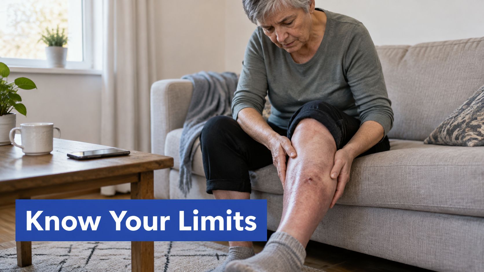
Home care has limits, and naming them is part of safe care. Rising infection signs or arterial warning signs need same-day in-person assessment whatever the distance to a clinic. A video call can review a wound photo, check bandage technique and adjust a plan. It cannot rule out deep infection, nor confirm that arterial supply will tolerate compression.
The access gap is documented. A recent qualitative study from rural Uganda found shortages of trained personnel, essential supplies and standardized treatment protocols in local health facilities, as described in this qualitative study of lymphedema care in a low-resource setting. A 2025 telehealth study from India offers a workable model for bridging it: remote follow-up covering limb examination, hygiene and wound care, elevation, exercises, compression bandages, suitable footwear, and discussion of how patients managed home care between visits.
The evidence that home-applied bandaging works is early but encouraging. A 2024 pilot study in Sri Lanka found that two weeks of short-stretch compression therapy reduced median limb volume by 11.3% among 10 participants with stage 3 filarial lymphedema, published in the American Journal of Tropical Medicine and Hygiene. Ten people is a small group, so treat the result as a signal that bandaging can be taught and delivered outside a specialist centre, not as a guarantee. Cost matters too: a 2024 community wound-service evaluation of 561 patients with lymphedema found mean per-patient costs fell from £1,457.10 to £964.40 after lymphedema specialists were embedded in community nursing and wound services, reported in the British Journal of Community Nursing.
Four Things to Ask For
- Ask who will look at the limb, whether that's a community nurse, a remote review, or an outreach clinic. Name the person and the interval.
- Ask who teaches bandage or garment application and how often the technique gets checked. A taught skill drifts without review.
- Keep the monitoring log so a remote reviewer can see the trend instead of a snapshot.
- Request a written plan covering compression format, dressings, measurement points, escalation signs and review dates, so any clinician who sees you can pick it up.
Sometimes the swelling has a venous source that won't yield to wraps and elevation. A minimally invasive procedure may be considered when venous disease causes persistent symptoms, or when compression, elevation and activity modification don't provide enough relief. That's a conversation worth having early when access to ongoing specialist care is hard to arrange. Vein & Wound Institute brings advanced vein care and specialized wound management together under one roof, which keeps the circulation question and the wound question in the same plan.
A wound on a swollen limb rarely closes because someone found the right dressing. It closes when the fluid, the arterial supply and the infection risk are each addressed in order, and when someone is watching the trend. Vein and Wound Institute treats lymphedema, chronic venous insufficiency, venous leg ulcers and other non-healing wounds across Los Angeles, Santa Barbara and Ventura Counties, combining vein evaluation, compression therapy, debridement and coordinated follow-up in one practice. If a wound on a swollen leg or arm has stalled, visit Vein and Wound Institute to have the circulation and the wound assessed together.