About 45.7% of people with lower-extremity lymphedema also have chronic venous insufficiency, so mixed venous-lymphatic disease is common and persistent leg swelling shouldn't automatically be blamed on veins alone. In practical terms, treating venous reflux may reduce pressure without fully restoring a lymphatic system that has been overloaded for years.
You may recognize the pattern. Your ankles swell by evening, your legs feel heavy, and the skin seems tighter or more irritated than it used to. Perhaps you've already been told that you have varicose veins, chronic venous insufficiency, or “just edema.” Treatment may have improved some symptoms, yet your shoes still feel tight and the swelling never completely disappears.
That experience doesn't necessarily mean treatment failed. It may mean that two drainage systems are involved. The veins return blood toward the heart, while the lymphatic vessels remove excess fluid and proteins from the tissues. When venous disease continues for long enough, the lymphatic system can become overwhelmed and damaged. Clinicians call this combined problem phlebolymphedema.
Table of Contents
- Why Your Leg Swelling Might Have Two Causes
- How Veins and Lymphatics Work Together
- Understanding Phlebolymphedema and Clinical Consensus
- Diagnostic Pathway for Mixed Venous-Lymphatic Disease
- Treatment Approaches for Combined Venous and Lymphatic Issues
- Preventing Progression Through Integrated Care
Why Your Leg Swelling Might Have Two Causes
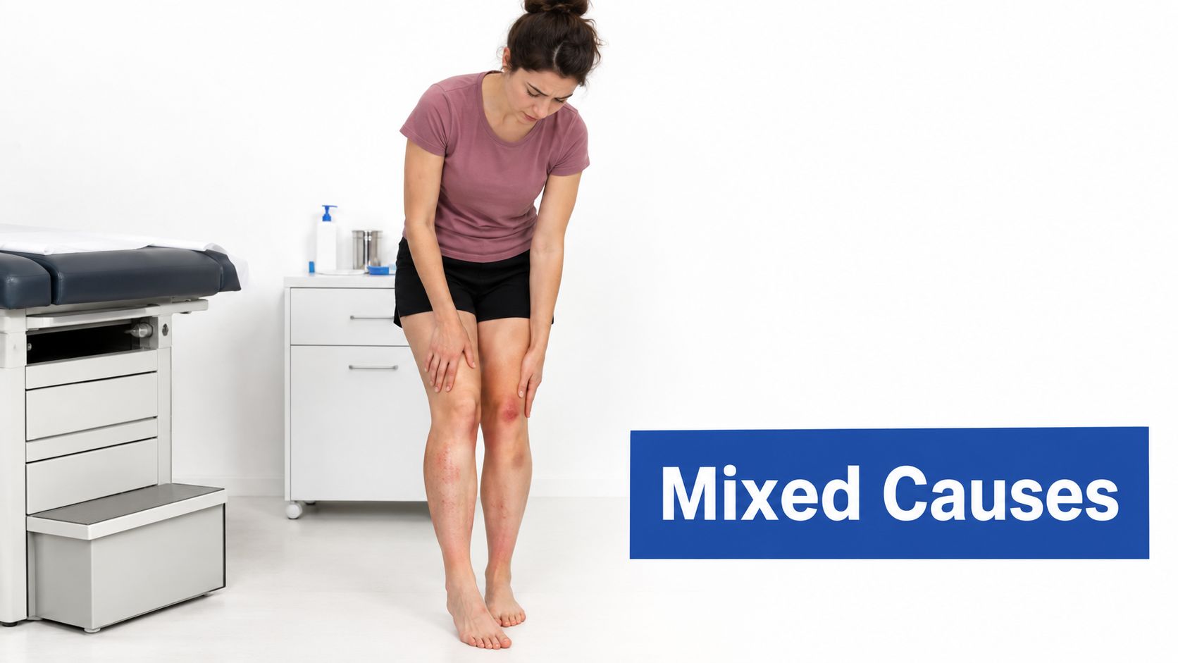
Consider a patient who has spent years standing at work and notices swelling around both ankles by the end of the day. A vein examination identifies reflux, meaning blood is moving backward because the valves aren't closing effectively. After treatment for the affected veins, the aching improves and the leg feels lighter, but the ankle remains enlarged and the skin feels firm.
That outcome can be confusing. The patient may reasonably think, “If the veins were the cause, why is the swelling still here?” The answer may be that venous pressure started the problem, but persistent fluid buildup has also impaired lymphatic drainage. The veins have received attention, while the second drainage pathway still needs care.
When improvement is incomplete
CVI-related swelling often begins as a fluid problem. It may be worse after prolonged sitting or standing and may ease with rest, movement, or elevation. Over time, however, repeated fluid overload can change the tissue itself. Skin may become thickened, irritated, or discolored, and the limb can feel less soft and more resistant to compression or elevation.
The overlap is clinically important. A 2026 meta-analysis found CVI in about 45.7% of people with lower-extremity lymphedema, with the estimate rising to 48.8% in studies using stricter definitions, although the researchers described the certainty as very low and the studies as highly variable (recent evidence on CVI and lower-extremity lymphedema). Those figures don't mean every swollen leg has both conditions, but they do show why a single-cause explanation can be incomplete.
Practical rule: If vein treatment improves pain or heaviness but swelling, tissue firmness, or skin problems persist, ask whether lymphatic dysfunction has also been assessed.
Persistent swelling also matters when wounds are present. A patient with lower-leg inflammation or an ulcer may need vascular evaluation alongside a structured wound plan. For background on the broader clinical checks used around diabetic wounds, diabetic foot ulcer assessment protocols can help explain why circulation, tissue condition, infection risk, and healing progress should be considered together.
A focused evaluation of lymphedema and its relationship to chronic swelling can help replace the unhelpful label of “just edema” with a more useful explanation of which systems need support.
How Veins and Lymphatics Work Together
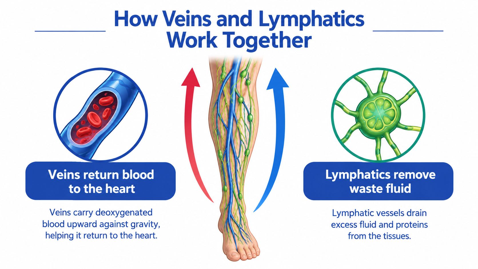
Think of the lower leg as a building with two drainage routes. The veins carry blood upward toward the heart, working against gravity with help from one-way valves and the calf-muscle pump. The lymphatic vessels collect fluid and proteins that leave the small blood vessels and return that material to the circulation through a separate network.
The two systems don't perform identical jobs, but they share the responsibility of keeping tissue fluid under control. If venous valves fail, blood pools in the lower limb and pressure rises inside the small vessels. More fluid then moves into the surrounding tissue. The lymphatic network initially increases its workload to remove the excess.
The drainage system under pressure
A useful analogy is a storm drain receiving water from a leaking roof. If the leak is small, the drain handles it. If the leak continues and the volume keeps increasing, the drain backs up, surrounding materials stay wet, and the drainage structure itself may deteriorate.
That is the mechanical link between CVI and lymphatic failure. Venous hypertension can push capillary filtration beyond the lymphatic system's transport capacity. At first, the lymphatics compensate. With persistent overload, they can become damaged, fluid clearance slows, and protein-rich fluid remains in the tissue. The body responds with inflammation and fibrosis, which makes the limb stiffer and harder to decompress. This progression is described in the clinical review of phlebolymphedema's venous and lymphatic mechanism.
CVI is widespread, although estimates vary because studies use different definitions and populations. In the United States, estimates place CVI at about 10% to 35% of adults, while reported prevalence varies by sex and age. The Edinburgh Vein Study found age-adjusted CVI in 9.4% of men and 6.6% of women, increasing to 21% of men over 50 and 12% of women over 50 (clinical overview of chronic venous insufficiency). The important point isn't one universal percentage. It's that venous disease becomes more common with age and can increase the workload placed on lymphatic drainage.
Why the tissue changes
Venous fluid is not the only concern. Lymphatic fluid contains proteins and cellular waste that need to leave the tissue. When those materials remain in place, the body can develop chronic inflammation, skin thickening, and fibrosis.
This explains why long-standing swelling may stop behaving like ordinary, temporary edema. Elevating the leg may help the fluid component, but it can't instantly reverse structural changes in the skin and connective tissue. A combined venous-lymphatic disorder therefore requires more than a short-term attempt to move fluid. It requires reducing the venous pressure and supporting lymphatic clearance over time.
Understanding Phlebolymphedema and Clinical Consensus
Phlebolymphedema means that venous disease and lymphatic dysfunction are contributing to the same swollen limb. In many cases, CVI comes first. Venous hypertension increases the amount of fluid entering the tissues, and the lymphatic system eventually can't remove it efficiently. The result is secondary lymphedema driven or amplified by venous disease.
This differs from primary lymphedema, in which the lymphatic system itself develops abnormally, and from swelling caused by a temporary injury or a systemic condition. The categories can overlap, so the diagnosis depends on the clinical pattern and the patient's history rather than on the word used to describe the swelling.
Why advanced venous disease changes the plan
Expert consensus from the American Venous Forum and American Vein and Lymphatic Society makes a significant practical recommendation: patients with CVI in CEAP classes C3 through C6 should be treated as lymphedema patients (consensus guidance for venous and lymphatic disease). These classes represent advanced clinical manifestations of venous disease, including edema, skin changes, and ulcers.
The recommendation changes the timing of care. Clinicians shouldn't wait until the limb becomes severely fibrotic or develops a wound before introducing lymphedema-oriented management. Compression, skin protection, movement, and ongoing monitoring belong in the plan when advanced CVI is present, even if venous reflux remains the most visible diagnosis.
Clinical examination is considered adequate for diagnosing lymphedema in this consensus. That doesn't mean every patient needs the same evaluation or that imaging is never useful. It means an experienced clinician can often identify the condition through the pattern of swelling, tissue texture, skin findings, and history.
Treating the patient rather than the label
A patient may hear “venous edema” and assume that a vein procedure will make every aspect of swelling disappear. Another patient may hear “lymphedema” and assume that the veins don't matter. Both assumptions can lead to incomplete care.
The more accurate question is, “How much of the swelling comes from venous pressure, and how much reflects impaired lymphatic transport?” A practical introduction to managing lymphedema with compression can help patients understand why compression remains relevant even after a venous intervention.
For a broader explanation of reflux, symptoms, and treatment decisions, the guide to chronic venous insufficiency symptoms, causes, and treatment provides useful context. The central clinical shift is simple: advanced CVI should trigger a combined care pathway rather than a vein-only pathway.
Diagnostic Pathway for Mixed Venous-Lymphatic Disease
A persistent swollen leg deserves a structured evaluation, especially when the swelling affects the foot or ankle, changes the skin, or doesn't resolve fully with elevation. Lower-limb lymphedema is often confused with CVI, deep vein thrombosis, or systemic edema. Recent literature describes diagnostic delays that can extend up to ten years, which is one reason early triage matters (clinical review of lower-limb lymphedema assessment).
Start with the whole pattern, not one symptom. A clinician should ask when the swelling began, whether it is one-sided or bilateral, how it changes during the day, whether the feet are involved, and whether there have been infections, wounds, cancer treatments, surgery, or clotting events.
A practical sequence
Review possible systemic causes. Heart, kidney, liver, medication-related, and other systemic explanations need appropriate medical assessment. Ruling out one category doesn't prove that the swelling is lymphatic, but it narrows the clinical picture.
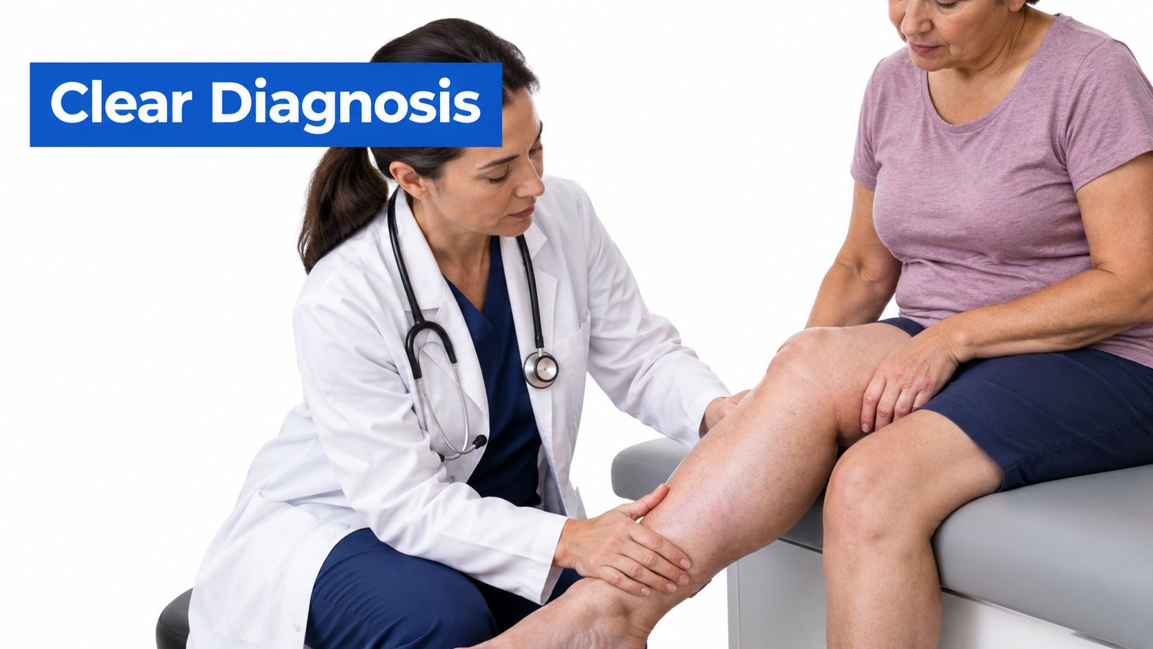
Examine the limb closely. The clinician should assess the feet, toes, ankles, calves, skin color, skin temperature, tissue firmness, wounds, and symmetry. Foot involvement and changes in tissue texture can raise suspicion for lymphatic dysfunction, while ankle staining, varicose veins, and dependent swelling may support venous disease.
Assess arterial safety before compression. An ankle-brachial index can help identify whether arterial circulation is adequate for the planned compression approach. Compression isn't a one-size-fits-all product, so the pressure and garment should match the vascular findings.
Check venous flow. Venous Doppler, often called duplex ultrasound, can identify reflux, obstruction, and abnormalities in deep or superficial veins. Visible varicose veins aren't required for venous disease, so a normal-looking skin surface shouldn't end the investigation.
Ask whether both systems need treatment. If chronic venous findings appear alongside persistent swelling, foot involvement, skin thickening, or a firm limb, the question shouldn't be “veins or lymphatics?” It should be whether the patient needs both vascular management and lymphedema-focused care.
Questions to bring to an appointment
Bring a short symptom record rather than relying on memory. Note whether swelling is worse after standing, whether it decreases overnight, which areas are affected, and whether the skin has become tight, rough, darker, or prone to leaking.
You can also ask:
- Could venous reflux be contributing even without large visible veins?
- Does the swelling pattern suggest lymphatic involvement?
- Is duplex ultrasound appropriate?
- Do I need an ankle-brachial index before using stronger compression?
- Should I see a clinician or therapist experienced in both venous disease and lymphedema?
Urgent assessment is appropriate for sudden one-sided swelling, new severe pain, chest symptoms, marked redness, fever, or a rapidly changing wound. Those features can signal problems that shouldn't be managed as routine chronic edema.
Treatment Approaches for Combined Venous and Lymphatic Issues
Treatment depends on which system is failing, how advanced the tissue changes are, and whether a wound or arterial concern is present. Compression remains foundational for both CVI and lymphedema, but the way it is selected and used matters. A garment that is too strong, poorly fitted, or unsafe for the arterial circulation can create problems rather than solve them.
A vein-focused plan may address reflux through an appropriate minimally invasive procedure, while a lymphatic-focused plan may include compression, skin care, exercise, and manual lymphatic drainage. When both conditions are present, those approaches work together rather than compete.
Comparing the main approaches
| Clinical situation | Main priority | Why it matters |
|---|---|---|
| Venous disease with mild, reversible swelling | Address venous pressure and use prescribed compression | Reducing reflux can decrease the amount of fluid entering the tissue |
| Persistent swelling after venous treatment | Continue compression and assess lymphatic function | Venous improvement may not reverse established lymphatic impairment |
| Firm tissue, foot involvement, or progressive shape changes | Add lymphedema-oriented therapy | Protein-rich fluid and fibrosis need more than elevation alone |
| Swelling with skin breakdown or an ulcer | Coordinate vascular and wound care | Healing depends on circulation, edema control, skin protection, and infection management |
Why vein treatment may not be enough
Correcting reflux can reduce one source of fluid overload. It can't guarantee that lymphatic vessels will immediately resume normal transport after prolonged stress. That's why some patients feel less aching after a venous procedure while the limb remains swollen or firm.
Compression helps limit new fluid accumulation and supports upward movement through the venous and lymphatic pathways. A clinician may recommend garments, bandaging, or a pneumatic device depending on the limb, the tissue, and the patient's ability to use the treatment consistently. The principles and practical considerations of compression therapy are especially relevant when swelling persists after the venous component has been addressed.
Additional support for advanced disease
Manual lymphatic drainage may be included when lymphatic dysfunction is significant, particularly as part of a broader decongestive program. It shouldn't be treated as a substitute for evaluating venous reflux or arterial circulation. Skin care also matters because stretched, dry, or cracked skin gives bacteria an easier route into the tissue.
Movement is another part of the plan. Walking and repeated ankle or toe movements activate the calf-muscle pump and can support fluid return. For a person who can't walk comfortably, a clinician can suggest safe, gradual exercises that fit the individual's condition.
Wounds require a separate level of attention. Compression may support healing in venous wounds, but the clinician must first consider arterial flow, infection, wound depth, tissue viability, and the cause of the ulcer. Treating the wound without addressing the edema often leaves the underlying pressure problem in place.
Preventing Progression Through Integrated Care
The best time to add lymphatic thinking is before the leg becomes rigid, repeatedly infected, or difficult to fit with compression. Once fibrosis and skin damage develop, care can still help, but the limb may respond more slowly and require more sustained management.
The burden of lymphedema is substantial. Global estimates commonly range from 140 million to 250 million affected people, with some contemporary summaries estimating about 10 million people in the United States (overview of lymphedema burden and causes). These figures include different forms of lymphedema, not only venous disease, but they reinforce why chronic swelling deserves more than casual reassurance.
A long-term prevention mindset
Integrated care means managing the cause and the consequence at the same time:
- Evaluate early: Persistent swelling, skin changes, or unexplained heaviness should prompt clinical assessment rather than repeated self-treatment.
- Protect the skin: Moisturize as appropriate, treat cracks promptly, and seek care for spreading redness, warmth, drainage, or fever.
- Use prescribed compression: Correct fit, pressure, and daily consistency matter more than buying a generic sock.
- Keep moving safely: Regular walking or clinician-approved ankle exercises support both venous return and lymphatic flow.
- Monitor after procedures: Improved veins don't eliminate the need to watch swelling, tissue texture, and wound healing.
Waiting for severe tissue changes before addressing lymphatic dysfunction is a poor strategy. Early evaluation preserves more options and gives the patient a clearer plan.
Patients with advanced CVI should be followed over time, even when symptoms improve. A coordinated clinician can reassess venous flow, compression needs, skin condition, and wound risk as the limb changes. That approach treats swelling as an ongoing vascular and lymphatic issue rather than a symptom that disappears once a single procedure is complete.
Vein and Wound Institute evaluates chronic venous insufficiency, leg swelling, lymphedema, and non-healing wounds with clinical examination and duplex ultrasound, then coordinates vein treatment, compression, and wound care when both systems are involved. If your swelling has persisted after treatment or you've been told it's “just edema,” visit Vein and Wound Institute to request an evaluation and discuss a plan for protecting your circulation and tissue health.