
You're putting on socks in the morning and notice it again. The skin just above your ankles looks reddish brown, a little shiny, and it itches more than it did last month. By evening your lower legs feel heavy and swollen, and the moisturizer you keep trying helps for a few hours, then stops helping. That pattern is common, and it's often not primarily a skin problem. When leg veins can't return blood upward efficiently, the skin is usually where the trouble becomes visible first.
Table of Contents
- Why Slower Leg Circulation Shows Up on the Skin
- How Venous Insufficiency Drives Skin Change
- Recognizing the Early and Advanced Signs
- Conditions That Look Similar and How to Tell Them Apart
- How Specialists Confirm the Diagnosis
- Treating the Vein and the Skin Together
- Making Compression Work in Real Life
- Long-Term Habits That Keep the Skin Stable
Why Slower Leg Circulation Shows Up on the Skin
A patient I see often describes the same sequence. It starts with itching around the inner ankle, then dry scaling that looks like eczema. The color shifts from pink to rusty brown. Swelling is modest in the morning and worse after a day of sitting or standing. Because the first complaint is skin, many people try antifungal creams, stronger moisturizers, or a short course of steroid ointment before anyone examines their veins.
That delay makes sense, but it misses the mechanism. Venous insufficiency stasis dermatitis is an inflammatory skin manifestation of chronic venous insufficiency, driven by sustained pressure inside leg veins that aren't moving blood upward well. In other words, the dermatitis is downstream evidence of an upstream circulation problem. Treating only the surface can calm itching temporarily while the pressure that caused it keeps building.
Why This Isn't a Cosmetic Issue
Age makes the pattern more likely, not less important. A narrative review notes approximately 2 to 6 million people in the United States have advanced chronic venous insufficiency, and stasis dermatitis was identified in 6.2% of people aged 65 years or older in one population study, as summarized in a 2023 narrative review of stasis dermatitis epidemiology and presentation. Comparable figures in older groups and a lower rate among younger people with varicose veins point to the same conclusion. This is an age-associated complication of chronic venous disease, not a cosmetic side effect of visible veins.
Several factors accumulate over time and raise risk:
- Prior leg clot or vein injury: Damage to valves or the vein wall makes backward flow more likely.
- Limited mobility: Calf muscles help pump blood upward, so immobility leaves more blood pooled in the lower leg.
- Obesity and chronic edema: Both increase the load the venous system has to manage.
- Long-standing varicose veins: Visible surface veins can signal deeper reflux that isn't obvious from the skin alone.
If lower leg skin changes keep returning despite reasonable skin care, ask what's happening underneath the skin, not just what cream to try next.
The practical reason to reframe the problem is timing. Dermatitis is an earlier, more reversible stage than hardened skin or an open ulcer. Recognizing it as a warning sign supports earlier duplex evaluation, a compression plan that is wearable, and treatment of reflux before fragile skin breaks down. Integrated vein and wound care follows that logic. It addresses the vein causing the pressure and the skin and tissue absorbing the damage, at the same time.
How Venous Insufficiency Drives Skin Change
Healthy leg veins have a simple job that's mechanically difficult. They carry blood from the feet back to the heart, against gravity, every time you stand. Small one-way valves inside the veins open as blood moves upward and close to stop it falling back down. Your calf muscles act as a pump when you walk, squeezing the deep veins and pushing blood toward the heart.
Problems start when valves weaken, vein walls stretch, or a prior clot scars the inside of a vein. Blood then flows backward, a pattern called reflux, and pools in the lower leg. If you've read about how chronic venous insufficiency develops and is treated, this pooling step is the turning point between a vein symptom and a skin disease.
From Pooled Blood to Inflamed Skin
Think of a slow drain in a sink. Water doesn't stop moving entirely, but it backs up, pressure rises, and eventually it starts leaking where it shouldn't. In the leg, sustained venous hypertension pushes fluid out of tiny capillaries into surrounding tissue. Red blood cells and inflammatory proteins leak out too, and the skin reacts to material that was never meant to sit there.
That sequence explains the classic distribution. Pressure is highest near the ankle, especially on the inner side, so edema, redness, scaling, and brownish discoloration cluster there first. As red blood cells break down in tissue, iron-containing pigment remains behind and produces the rusty hyperpigmentation patients often mistake for bruising that won't fade.
The infographic below maps that progression from valve failure to skin breakdown.
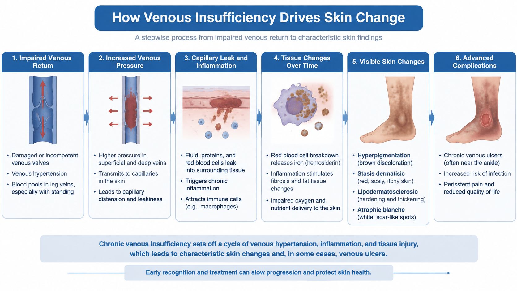
Early disease is mainly inflammatory. The skin is itchy, red, and swollen, but it's still relatively soft and responsive when pressure is reduced. Later disease becomes fibrotic. Chronic inflammation stimulates scar-like change in skin and fat, producing lipodermatosclerosis, where tissue feels woody and tight and the lower leg can narrow above the ankle. At that stage the skin tolerates minor trauma poorly, and a small scratch or bump can become a venous ulcer that heals slowly.
That's why skin-directed treatment alone rarely holds. A steroid can quiet inflammation and an emollient can reduce cracking, but neither lowers venous pressure. Until reflux, obstruction, and edema are addressed, the same leakage keeps restarting the cycle.
Recognizing the Early and Advanced Signs
Stasis dermatitis usually develops gradually, and people often adapt to each small change until the leg looks quite different from a year earlier. Comparing both legs, checking how symptoms change through the day, and noticing whether elevation helps can give you useful information to bring to an appointment.
Early Changes People Often Dismiss
Early signs tend to be sensory before they're dramatic visually. Itching is frequently the dominant complaint, sometimes with burning or a tight feeling after standing. You may see patchy redness, fine scaling, and dryness around the ankle that resembles ordinary eczema. Edema at this stage often pits, meaning a fingertip pressed into the swelling leaves a temporary dent, and it's typically worse in the evening than on waking.
Look for this early cluster:
- Itch more than pain: Persistent itching, irritation, or sensitivity to socks and creams.
- Color change near the ankle: Pink, red, or faint brown patches, often on the inner leg.
- Dry, scaly texture: Flaking, crusting, or small cracks that sting in the shower.
- Dependent swelling: Puffiness that builds during the day and eases overnight or with elevation.
Signs the Disease Is Advancing
As venous pressure persists, pigment deepens into a more uniform brown or reddish brown discoloration that doesn't blanch when pressed. Skin can look shiny because it's stretched by edema, then progressively thicker. Induration, a firm, hardened quality under the surface, signals that inflammation is moving toward fibrosis. In lipodermatosclerosis the tissue feels woody, tenderness is common, and the leg may take on a narrowed shape above the ankle.
Other warning signs deserve prompt attention. Small white, scar-like patches can appear within darker skin and indicate areas of poor local blood supply and fragility. Weeping, crusting, or recurrent superficial cracks mean the barrier is failing. Any open area that isn't closing, especially just above the inner ankle, should be treated as a possible early venous ulcer rather than a routine scrape.
Practical rule: Itchy, discolored skin plus swelling that worsens through the day deserves a vein evaluation, even if the skin hasn't broken open.
You don't need to stage yourself precisely at home. The useful distinction is simpler. Mild, stable dryness without swelling is different from persistent dermatitis with pigment change, hardening, weeping, or an open sore. Once you're in that second group, watchful waiting with moisturizer alone usually loses ground.
Conditions That Look Similar and How to Tell Them Apart
Red lower legs get mislabeled often, and the label matters because treatment diverges quickly. Stasis dermatitis needs venous management and usually compression. Cellulitis may need urgent antimicrobial treatment. Significant arterial disease can make standard compression unsafe. Getting the category wrong in either direction causes harm, either from unnecessary antibiotics and missed vein disease, or from delayed treatment of a real infection.
The framework I use with patients starts with a few discriminating questions. Is it one leg or both? Does it itch or hurt? Did it develop over months or over a day or two? Is there fever, drainage, or rapid spread? Those answers don't replace an exam, but they change urgency. This diagnostic overlap, including contact dermatitis developing on top of venous disease, is addressed in the Merck Manual overview of stasis dermatitis and its mimics.
Clues That Point Away From Simple Stasis Dermatitis
Cellulitis usually affects one leg, comes on relatively quickly, and hurts more than it itches. Warmth, expanding redness, fever, chills, streaking, or feeling unwell push strongly toward infection and same-day evaluation. Stasis dermatitis, by contrast, is often bilateral, chronic, itch-dominant, and accompanied by pigment change and varicose veins. The trap is that an acute flare of stasis dermatitis can look angry enough to be mistaken for cellulitis, and true cellulitis can also develop in a leg that already has stasis changes.
Contact dermatitis is the other frequent confounder, and it can coexist with stasis dermatitis rather than replace it. A new dressing adhesive, topical antibiotic, fragranced emollient, or compression material can trigger itching and redness that maps onto where the product touches the skin. If symptoms sharpen right after a product change, or stay sharply limited to a bandage outline, contact irritation or allergy moves up the list.
The chart below is included to prompt structured symptom comparison before you decide how urgent a change feels.
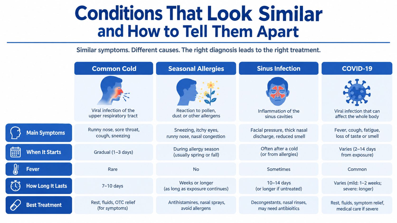
Lymphedema tends to involve the foot and toes more prominently and produces swelling that becomes less pitting over time, with thickened skin folds. It can overlap with venous disease, which is why some patients need treatment for both systems. Arterial insufficiency points in a different direction, with cool skin, weak or absent pulses, pain that's worse with elevation, and ulcers more often on the toes, heel, or outer leg. Ulcer location and pain pattern are especially useful, as explained in this comparison of venous and arterial leg ulcers.
Seek care urgently for rapidly spreading redness, fever, severe or escalating pain, new drainage with odor, a suddenly much more swollen leg, or skin turning pale, blue, or cold. Those features should not be managed by tightening compression alone and waiting.
How Specialists Confirm the Diagnosis
A first visit for suspected stasis dermatitis is mostly noninvasive, but it shouldn't be superficial. I want to know how long the skin changes have been present, whether they're new or recurrent, what products you've applied, and whether you've had a leg clot, vein procedure, injury, or ulcer before. Medication history, diabetes, heart or kidney disease, mobility limits, and prior compression use all change the plan.
What the Exam and Ultrasound Show
On examination, the clinician looks at distribution and texture of the dermatitis, edema, varicose veins, pigment, induration, and any open areas. Pulses at the foot are checked, sensation is assessed when diabetes is present, and both legs are compared. A bilateral, itchy, pigmented pattern around the ankles in a leg with varicosities tells a different story than a unilateral, hot, painful leg that changed overnight.
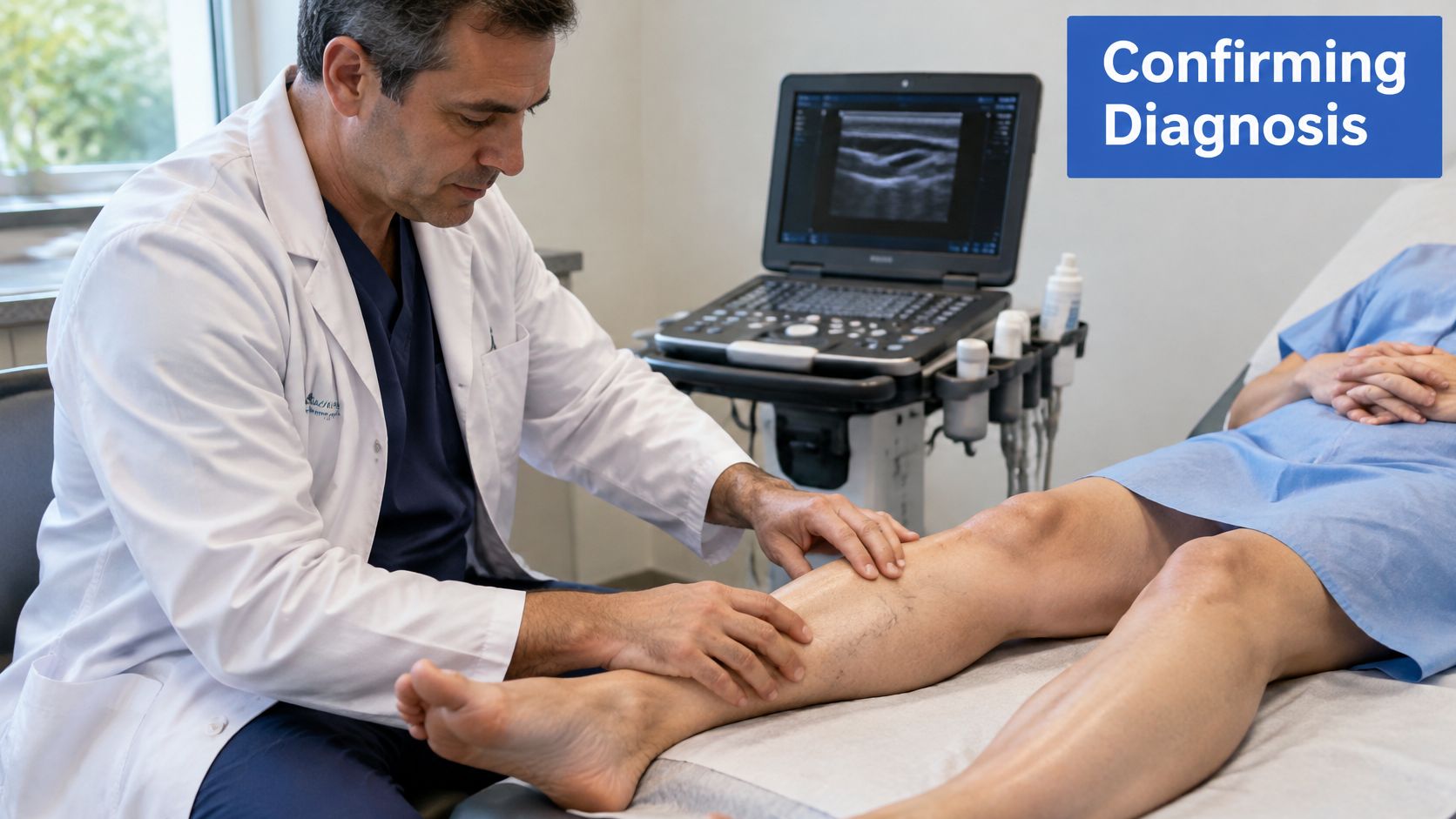
Duplex ultrasound is the central test for the venous side. It shows vein anatomy and blood flow in real time, identifies reflux through incompetent valves, and can reveal obstruction or evidence of prior thrombosis. That map determines whether compression alone is reasonable, which veins might benefit from a procedure, and whether another cause of swelling needs attention.
The image below shows the kind of ultrasound-based leg examination used to assess venous flow in clinic.
Why Arterial Testing Comes Before Compression
Compression shouldn't start blindly when arterial disease is possible. An ankle-brachial index or another perfusion assessment compares blood pressure at the ankle and arm and helps judge whether the leg can tolerate external pressure. Results need context, particularly with diabetes or calcified arteries, where readings can mislead and additional testing may be needed. If ischemia is suspected, compression is modified or avoided until arterial status is clarified.
Blood work isn't required for every classic case. It's ordered when the picture is atypical, infection is a concern, healing is poor, or another systemic condition may be contributing. If you're reviewing results from that kind of workup, a plain-language walkthrough of lab results explained step by step can help you understand what each value means before your follow-up conversation. Skin biopsy is uncommon, reserved for presentations that don't fit, such as possible vasculitis, atypical infection, or a lesion that looks unlike venous disease.
Bring a list of every cream, dressing, and supplement you're using, plus photos showing how the leg has changed. That history often identifies a contact trigger or a failed compression attempt that the exam alone won't reveal.
Treating the Vein and the Skin Together
Effective treatment has two targets that shouldn't be sequenced months apart. You have to lower venous pressure and control edema, and you have to repair and protect skin that's already inflamed or broken. Doing only one half explains many recurrences. A vein procedure won't instantly heal a colonized, macerated ulcer, and excellent wound dressings won't keep an ulcer closed if reflux keeps driving pressure into the same tissue.
Lowering Pressure and Treating Reflux
Compression is the principal pressure-based intervention because it narrows distended veins, improves venous return, limits dependent edema, and reduces ambulatory venous pressure. For venous leg ulcers, a clinical review reports typical targets of approximately 30 to 40 mmHg and notes that multicomponent systems generally heal ulcers more effectively than single-component systems, as detailed in a clinical review of compression therapy for venous leg ulcers. Without an ulcer, pressure and format are individualized to swelling, limb shape, mobility, skin tolerance, and your ability to apply the garment.
When duplex shows treatable reflux, closing or removing the faulty vein can reduce the pressure source. Options include endovenous laser ablation and radiofrequency ablation, which use heat inside the vein, adhesive closure with VenaSeal, mechanochemical treatment with ClariVein, and foam-based approaches including Varithena and ultrasound-guided foam sclerotherapy. Bulging surface tributaries may be removed through tiny incisions with ambulatory phlebectomy. These aren't interchangeable menu items. Vein diameter and depth, anatomy near nerves, prior clot, skin condition, and whether an ulcer is active all influence which approach, if any, fits.
Treating reflux doesn't replace compression during healing. In most active dermatitis and ulcer care, they work as a pair.
Calming Skin and Healing Open Tissue
Skin care during a flare is deliberately plain. Gentle cleansing, liberal bland emollients, and a time-limited topical corticosteroid when prescribed can reduce itching and cracking. Products with fragrance or a long ingredient list create avoidable contact dermatitis risk on already reactive skin. Scratching feels harmless and isn't. On edematous, pigmented skin it creates entry points for bacteria and small erosions that expand.
If an ulcer or non-healing wound is present, care escalates beyond surface dermatitis management. Excisional debridement removes dead tissue that stalls healing, advanced dressings manage drainage and protect surrounding skin, and infection is treated based on clinical signs rather than the mere presence of bacteria on a surface swab. For wounds that stall despite pressure control and good local care, bioengineered skin substitutes and grafts may be used to support closure. The sequence matters. A graft placed into uncontrolled edema and untreated reflux is being asked to survive in the same environment that created the wound.
Persistent dermatitis or recurrent ulceration despite reliable compression isn't a reason to just try harder with the same plan. It warrants reassessment for untreated reflux or obstruction, infection, a contact trigger, arterial limitation, or compression that isn't delivering the intended pressure because of fit or application technique.
Making Compression Work in Real Life
Telling someone to wear compression is easy. Getting compression onto a swollen, tender leg every morning, with arthritic hands, limited ability to bend, and a dressing that needs to stay in place, is the actual problem. Adherence fails most often because of discomfort, donning difficulty, heat, cost, and garments that no longer fit after swelling changes. Those are solvable engineering and behavior problems, not character flaws.
Match the Device to the Obstacle
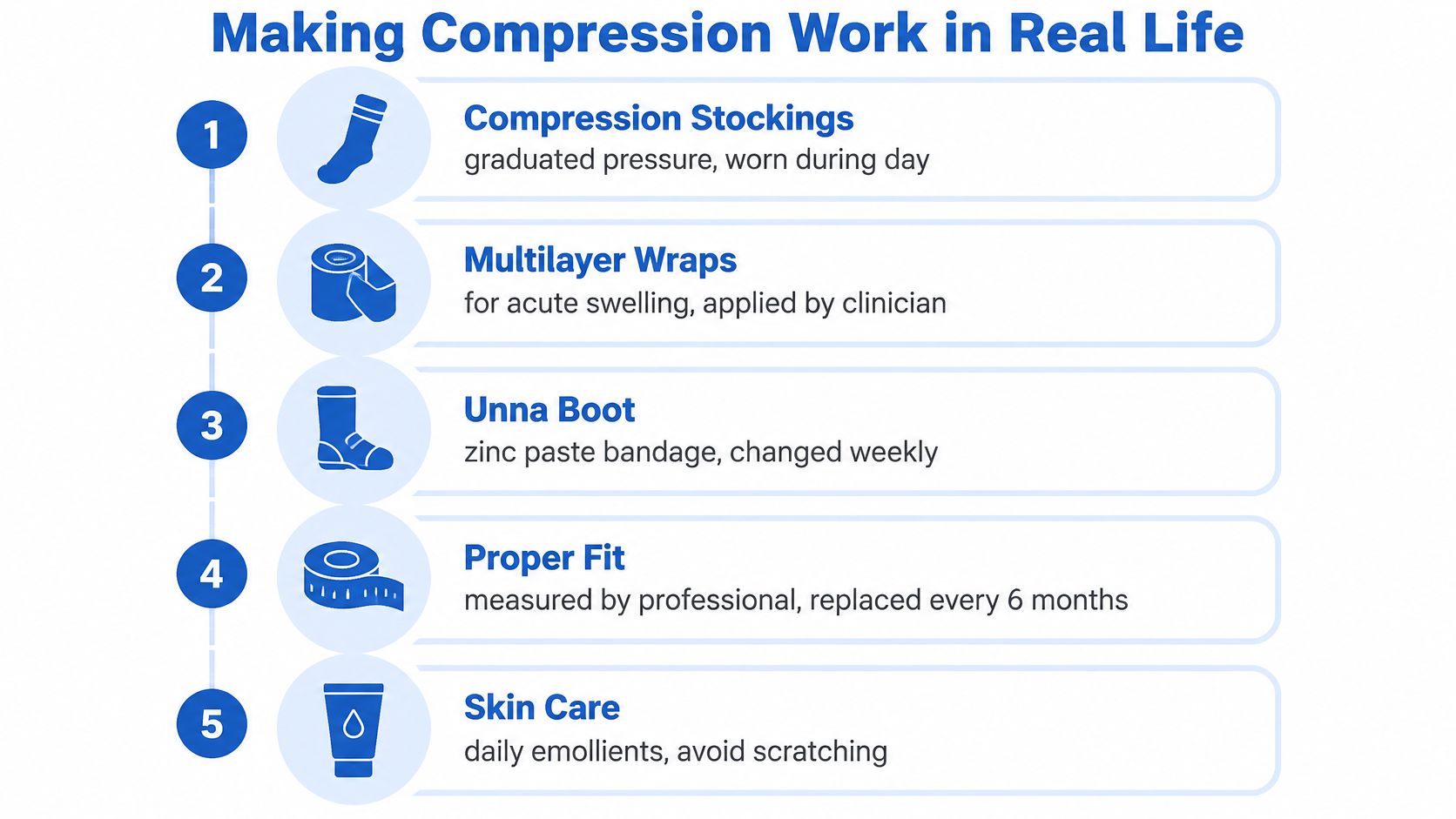
Graduated stockings suit many people with stable edema and intact skin who can apply them reliably. They work poorly when leg shape changes quickly, drainage soaks fabric, or grip strength is limited. In those situations, adjustable multilayer wraps applied by a clinician, or systems you can tighten with straps rather than pull over the heel, may deliver more consistent pressure. An Unna boot, a zinc paste bandage system, can be useful when dermatitis and light drainage need protection between scheduled changes and daily self-application isn't realistic.
For patients who can't tolerate standard approaches, pneumatic options are being studied as adjuncts rather than universal replacements. A 2025 randomized proof-of-concept trial found adding intermittent pneumatic impulse compression to standard treatment significantly reduced ankle edema and improved tissue oxygenation over five days, while elastic wrapping alone didn't show a significant change in that study, as reported in a randomized proof-of-concept trial of pneumatic impulse compression. That's promising, but its single-center proof-of-concept design limits generalization. It doesn't retire fitted compression, and it doesn't remove the need for arterial assessment first.
Details on formats and clinical use are outlined on the compression therapy service page, but the selection logic in clinic is practical. Severe swelling needs adjustability, active drainage needs a system compatible with dressings, and poor hand strength needs donning aids, gloves for grip, or a wrap design that doesn't depend on pulling.
The checklist below summarizes the main options and the daily habits that make them work.
Daily Technique and Troubleshooting
Apply compression first thing in the morning, before dependent swelling builds, on clean, moisturized, fully dry skin. Smooth out wrinkles, because a folded band creates a tourniquet-like ridge and a pressure sore. Check toes and skin daily for color change, new numbness, blistering, or increasing pain. Those symptoms mean remove it and contact the clinic rather than pushing through.
When standard stockings fail, work through the cause:
- Can't get them on: Try a donning frame or gloves, or switch to an adjustable wrap you can secure without pulling over the heel.
- They roll or dig in: Limb shape has likely changed, so get remeasured rather than folding the top band down.
- Skin gets itchier underneath: Review detergents, emollients, and garment materials for a contact trigger.
- Swelling isn't improving: Wear time, fit, elasticity loss, and untreated reflux all need review, not just more willpower.
Garments lose elasticity with use and washing, so a stocking that once worked can stop delivering useful pressure. Track simple follow-up measures that show whether the plan is working, including ankle swelling, daily wear time, skin comfort, and ulcer size if one is present.
Long-Term Habits That Keep the Skin Stable
Once dermatitis quiets, the goal shifts from rescue to maintenance. Walk daily if you're able, because calf muscle contraction is your built-in venous pump, and break up long periods of sitting or standing with movement. Raise your legs above heart level during rest periods to drain dependent edema rather than letting it accumulate until bedtime.
Keep skin care boring and consistent. Use bland emollients daily, avoid fragranced products and unnecessary topical antibiotics, and protect pigmented, thinned skin from bumps, scratching, and tight footwear. Weight management and treatment of conditions that worsen edema support the venous plan, but they don't substitute for compression or follow-up duplex checks when symptoms change.
At the first sign of recurrence, meaning returning itch, scaling, new pigment, or swelling that's no longer controlled, restart the conversation early rather than waiting for a crack or ulcer. Rapidly spreading redness, fever, escalating pain, drainage, or a pale, cold foot needs urgent assessment. Stable skin over years is a realistic goal for many people, but it's maintained, not cured once and forgotten.
If itchy, discolored, swollen lower legs haven't responded to skin care alone, an integrated evaluation can assess your venous circulation with duplex ultrasound, check arterial status before compression, and build a combined vein, skin, and wound plan around what your legs need. Visit Vein and Wound Institute to learn about evaluation and treatment options and to schedule a consultation.