Chronic venous insufficiency can start with something as ordinary as end-of-day ankle swelling or heavy legs, yet it may progress to permanent skin changes and open wounds. In the United States, an estimated 10% to 35% of adults have chronic venous insufficiency, and the condition becomes more common with age according to the National Center for Biotechnology Information. Visible veins aren't the only clue. Positional swelling, aching, itching, brown discoloration, hardened tissue, and wounds can all reflect impaired venous return and reflux.
The challenge is that these symptoms overlap with lymphedema, arterial disease, neuropathy, thrombosis, and other conditions. The eight signs below move from earlier, often reversible symptoms toward more serious tissue damage. Track when symptoms appear, whether elevation changes them, and whether one leg differs from the other. Clinician-directed compression, movement, skin care, and a vascular assessment may all have a role. A duplex ultrasound can help clinicians evaluate blood flow, valve function, and venous reflux, but this article can't diagnose the cause of your symptoms.
Table of Contents
- 1. Edema or Leg Swelling
- 2. Venous Claudication and Leg Heaviness
- 3. Varicose Veins
- 4. Telangiectasia and Spider Veins
- 5. Skin Discoloration and Pigmentation Changes
- 6. Lipodermatosclerosis
- 7. Atrophie Blanche
- 8. Venous Ulcers or Open Wounds
- 8-Point Comparison of Venous Insufficiency Signs in Legs
- Know When a Sign Needs Evaluation
1. Edema or Leg Swelling
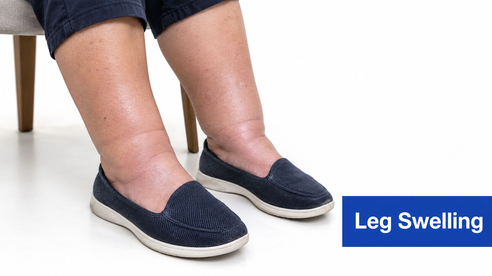
Swelling is often the first noticeable sign of venous insufficiency in legs. It usually appears around the ankles and lower calves, where gravity places the greatest demand on the veins. As weakened or damaged valves allow blood to move backward and pool, pressure rises in the leg veins and fluid can move into surrounding tissue.
A typical pattern is less swelling after overnight rest and more swelling by late afternoon or evening. Shoes, socks, or trouser cuffs may feel tighter as the day progresses. Some people develop swelling in both legs, while others have more noticeable edema on one side, particularly after previous vein injury or thrombosis.
What the pattern can tell you
Venous edema commonly worsens with prolonged standing or sitting and improves with leg elevation or compression, a pattern described in clinical guidance from the American Heart Association journal Circulation. That pattern supports a venous contribution, but it doesn't prove one. Heart, kidney, medication-related, lymphatic, and clot-related causes can also produce swelling.
Pay attention to the details:
- Timing: Record whether swelling is mild in the morning and more pronounced at night.
- Distribution: Note whether it affects the ankle, calf, or entire leg.
- Symmetry: Marked difference between legs deserves clinical attention.
- Response: Record whether elevation reduces the swelling or only provides temporary relief.
Don't begin firm compression without professional guidance if the cause of swelling is unclear. A clinician may need to assess circulation and use duplex ultrasound when thrombosis or another vascular problem must be considered. In confirmed venous disease, a plan may combine appropriately fitted compression, regular walking, elevation, and treatment of the underlying reflux. Salt reduction may help some people with fluid retention, but it won't repair faulty venous valves.
2. Venous Claudication and Leg Heaviness
Some patients don't describe swelling first. They describe legs that feel heavy, full, tired, or pressurized, especially after standing through a workday or sitting for a long period. The discomfort may be dull rather than sharp, and the legs can feel increasingly difficult to use as the day goes on.
Venous heaviness relates to pressure and congestion in the lower limbs. Edema can add to the sensation by making the tissues feel tight. A person might say that the legs feel as though fluid is collecting in them, or that walking becomes less comfortable even though there is no obvious injury.
Position matters
The most useful observation is what changes the symptom. Venous discomfort often worsens while standing or walking and improves with rest, lying down, or elevation. The Cleveland Clinic's clinical overview of chronic venous insufficiency also describes heaviness, aching, fatigue, itching, burning, tingling, and swelling as possible symptoms, sometimes before prominent veins appear.
That pattern differs from some forms of arterial disease, where pain during walking that improves with rest may reflect reduced arterial blood flow. The distinction isn't reliable enough for self-diagnosis, however. Restless-legs-like sensations, nerve problems, musculoskeletal pain, and venous disease can overlap. Some superficial venous disease may produce few symptoms, while deeper venous problems may cause more discomfort, so the presence or absence of pain doesn't settle the question.
A practical approach is to keep a short symptom record. Note how long you can stand, whether a break with elevation helps, and whether heaviness interferes with work, exercise, or sleep. Clinician-directed compression and regular calf-muscle activity may reduce venous pooling for some patients. If symptoms persist, worsen, or occur mainly in one leg, arrange a vascular evaluation rather than treating the sensation as simple fatigue.
3. Varicose Veins
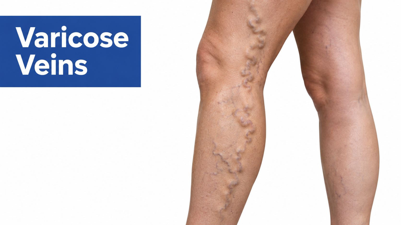
Varicose veins are enlarged, twisted, or rope-like veins visible beneath the skin. They develop when venous valves don't keep blood moving efficiently toward the heart. Reflux increases pressure inside the affected veins, which can stretch their walls and make them more prominent.
Some varicose veins are mainly a cosmetic concern. Others cause aching, heaviness, itching, cramps, or tenderness. Their appearance can also provide a visible clue to a deeper circulation problem, especially when they occur alongside swelling or symptoms that worsen with standing.
Appearance isn't the whole assessment
A vein's size or visibility doesn't reliably show how much reflux is present. Duplex ultrasound can map the affected veins and help clinicians determine whether symptoms come from superficial reflux, deeper venous disease, or another cause. That information matters before choosing an intervention.
Conservative measures can help manage symptoms, but they won't eliminate established reflux. Movement activates the calf-muscle pump, elevation can reduce pressure temporarily, and clinician-selected compression may make standing more comfortable. Treatment decisions depend on the vein pattern and the person's symptoms. Options may include endovenous procedures, foam sclerotherapy, or removal of selected surface veins, but the appropriate choice requires imaging and examination.
Practical rule: Treat the circulation problem, not just the visible vein. Surface treatment without understanding the underlying reflux can leave the main source of symptoms unaddressed.
Patients often benefit from learning how to prevent varicose veins and maintain healthy legs, particularly when they spend long periods standing or sitting. Seek assessment sooner if varicose veins occur with persistent swelling, skin discoloration, tenderness, or a sore near the ankle. Those additional findings suggest that the issue may have progressed beyond an isolated cosmetic concern.
4. Telangiectasia and Spider Veins
Spider veins are fine red, blue, or purple lines that form web-like or branching patterns close to the skin. Clinicians may also call them telangiectasia, meaning small dilated vessels that have become visible. They commonly appear on the thighs and lower legs and may be asymptomatic.
A small cluster can be primarily superficial and cosmetic. In other cases, spider veins develop in association with reflux in a larger vein beneath the skin. Mild burning, itching, or aching makes a circulation assessment more useful, particularly when the veins are concentrated around the lower leg or occur with swelling.
When a cosmetic sign deserves imaging
The right question isn't "How many spider veins are there?" It is whether a larger refluxing vein is feeding them. Duplex ultrasound can help distinguish an isolated superficial pattern from telangiectasia associated with venous reflux. If reflux is present, clinicians may address that source before treating residual surface vessels.
Sclerotherapy can be useful for selected spider veins and small varicose tributaries. It involves injecting a solution that irritates the vessel so the body can close and gradually clear it. The result depends on the vessel pattern, the underlying circulation, and the patient's healing response. Some visible veins may remain or new ones may develop, so expectations should be realistic.
The benefits of sclerotherapy for spider veins and varicose veins are best considered after a clinician determines whether the veins are isolated or connected to larger refluxing segments. Compression may be recommended after treatment, but the garment and duration should be selected for the individual. Don't assume that every spider vein requires a procedure, and don't assume that every cluster is purely cosmetic.
5. Skin Discoloration and Pigmentation Changes
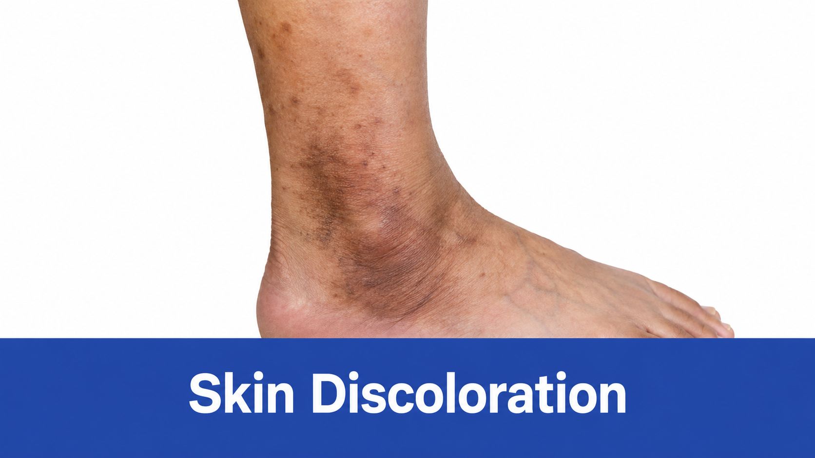
Brown, rust-colored, or reddish skin around the ankle and lower leg is a more meaningful warning sign than many people realize. Chronic venous hypertension can allow red blood cells to escape from small vessels into nearby tissue. As blood products break down, hemosiderin can accumulate and stain the skin.
This discoloration often appears on the inner ankle or lower calf. It may start subtly, then become more obvious over time. It can occur with itching, dryness, eczema-like irritation, swelling, or changes in skin texture. A person may mistake the color for bruising, sun exposure, or an old injury, especially when the process is gradual.
Why pigmentation changes the conversation
The CEAP system classifies edema as C3 and adds skin damage such as pigmentation, eczema, lipodermatosclerosis, or atrophie blanche under C4. That distinction matters because persistent skin change indicates progression beyond uncomplicated visible veins, as described in the Circulation review of chronic venous disease.
Existing discoloration may not disappear quickly, even when treatment improves venous circulation. The practical goal is to prevent further tissue damage, protect the skin barrier, and identify reflux that may be treatable. A clinician can examine for dermatitis, hardening, fragile areas, or an early wound.
Take a photograph in consistent lighting if you're monitoring change, and avoid scratching dry or itchy skin. Use a clinician-recommended moisturizer, protect the area from trauma, and ask whether compression is safe and appropriate. Sudden redness, warmth, rapidly increasing pain, or drainage needs prompt evaluation because infection or another acute problem may be present.
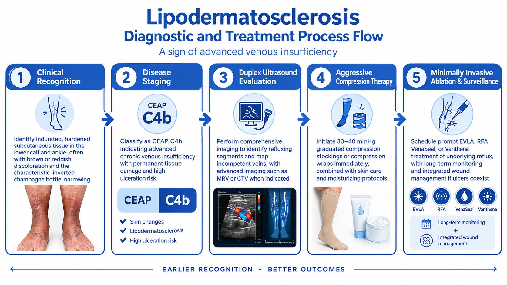
6. Lipodermatosclerosis
Lipodermatosclerosis is a hardened, inflamed change in the lower leg's skin and underlying tissue. The area may feel firm, tight, or painful rather than soft and pliable. It often appears with brown or reddish discoloration and can create a narrowed lower calf above a broader ankle, sometimes described as an inverted champagne-bottle shape.
This isn't just dry skin or an old varicose vein. It reflects long-standing venous pressure and inflammation that have altered the tissue. In CEAP classification, lipodermatosclerosis is an advanced C4 skin finding. It signals that the leg needs more than cosmetic management.
Protecting hardened tissue
The first practical step is a complete assessment. A clinician may evaluate the veins with duplex ultrasound, examine the skin for dermatitis or infection, and consider whether arterial circulation is adequate for compression. Treatment often focuses on improving venous return, protecting the skin, reducing edema, and addressing reflux when appropriate.
Compression can be helpful, but stronger is not automatically better. A clinician must select the pressure, garment, or wrap based on circulation, limb shape, swelling, comfort, and the person's ability to apply it. Poorly fitted compression can cause pressure points, discourage adherence, or damage vulnerable skin.
Hardened skin is a reason to arrange assessment, not a reason to massage aggressively or wait for an ulcer.
Moisturizing helps reduce cracking, but lotion alone won't reverse venous hypertension. Check the leg regularly for blisters, fissures, tenderness, drainage, or a new open area. People with diabetes, reduced mobility, prior thrombosis, or previous ulcers may need coordinated monitoring because several factors can slow healing at the same time.
7. Atrophie Blanche
Atrophie blanche appears as small, pale or white areas of thin, scar-like skin, often on the ankle or lower leg. The patches may occur among brown pigmentation, hardened tissue, or fine surrounding vessels. The affected skin has lost some of its normal structure and can be more vulnerable to minor trauma.
This sign indicates substantial chronic tissue change. It isn't the same as ordinary dry skin, a temporary pressure mark, or a harmless pale spot. Because it may occur near other advanced venous findings, clinicians should examine the entire lower leg rather than focusing only on the white macules.
Fragile skin needs a prevention plan
Atrophie blanche can be painful, particularly if the surrounding tissue is inflamed or under pressure. A person may avoid walking or compression because the area feels tender, but reduced movement can worsen venous pooling. The care plan therefore has to balance circulation support with skin protection and tolerability.
Protect the area from rubbing, sharp edges, and poorly fitting footwear. Padding may help reduce trauma, while gentle cleansing and regular moisturization support the skin barrier. Compression may be appropriate, but only after a clinician has assessed arterial circulation and selected a safe, workable method.
A specialist may also look for active dermatitis, infection, a healed ulcer, or a developing wound hidden among the scarred areas. Keep follow-up appointments if the leg has a history of ulceration or recurrent skin breakdown. Early attention to a new crack, blister, bleeding point, or drainage is more useful than waiting for a larger wound.
The appearance of atrophie blanche doesn't identify the exact cause by itself. Venous disease, inflammatory conditions, and other disorders can produce overlapping skin findings. A coordinated vascular and dermatologic assessment is appropriate when the diagnosis is uncertain, the lesions are painful, or the surrounding skin is changing.
8. Venous Ulcers or Open Wounds
An open sore near the inner ankle or lower leg is the most serious sign on this list. Venous leg ulcers develop when chronic venous hypertension, inflammation, edema, and impaired microcirculation prevent damaged tissue from healing normally. They often have irregular borders and may produce drainage, but appearance alone can't confirm that a wound is venous.
Venous leg ulcers are the most advanced clinically important sign of venous insufficiency. They account for the majority of chronic lower-extremity ulcers, and one review found that venous insufficiency causes nearly 80% of all lower-leg ulcer cases in the referenced review. The same source describes venous ulcers as an important consequence of progression from edema and skin changes to chronic wound disease.
Why wound and vein care belong together
Dressings can manage drainage and protect the wound, but they don't correct reflux. A thorough evaluation may include duplex ultrasound and an ankle-brachial index or another arterial evaluation, particularly before applying substantial compression. Arterial disease, diabetes, pressure, infection, neuropathy, and lymphatic problems can coexist with venous insufficiency and change the treatment plan.
Compression is central to many venous-ulcer plans, but the type and pressure must be prescribed after circulation has been evaluated. Wound care may include cleansing, appropriate dressings, debridement, infection management, and protection of the surrounding skin. When reflux contributes to the wound, treating the underlying vein problem may be considered alongside wound treatment.
Read more about the key differences between venous leg ulcers and arterial leg ulcers before assuming that every ankle wound has the same cause. Seek prompt medical attention for increasing pain, spreading redness, warmth, pus-like drainage, fever, or a wound that is enlarging or not progressing toward healing. An open wound shouldn't be managed solely with over-the-counter creams or a borrowed compression garment.
8-Point Comparison of Venous Insufficiency Signs in Legs
| Sign / Symptom | Typical presentation | Diagnostic & resource requirements | Implementation complexity | Expected outcomes | Ideal use cases / Key advantages |
|---|---|---|---|---|---|
| Edema (Leg Swelling) | Bilateral ankle/calf swelling; worse by day; pitting early → non‑pitting chronic | Clinical exam; leg circumference; duplex US to assess reflux/DVT; basic labs if systemic causes suspected | Low–moderate: elevation, compression, lifestyle; ablation if reflux present | Often improves quickly with compression/elevation; reversible early; can become fibrotic if chronic | Early CVI detection and monitoring; objective, easily measured sign guiding therapy |
| Venous Claudication & Leg Heaviness | Heavy, full, aching legs with prolonged standing/walking; rapid relief with elevation | Symptom scoring, clinical exam, duplex US to correlate reflux | Low–moderate: conservative (compression, exercise) → ablation for symptomatic reflux | Good symptomatic relief within weeks after appropriate ablation; subjective but measurable | Symptoms driving treatment decisions; improves function and quality of life |
| Varicose Veins | Visible tortuous veins >3 mm; may cause pain, itching, cosmetic concern | Clinical inspection and duplex mapping of saphenous/tributary reflux | Moderate: EVLA/RFA, sclerotherapy, phlebectomy; may need staged sessions | High symptomatic and cosmetic improvement; recurrence possible if reflux untreated | Symptomatic or cosmetic cases with documented reflux; multiple minimally invasive options |
| Telangiectasia & Spider Veins | Fine red/purple branching lines (<1–3 mm); often cosmetic; may itch/burn | Clinical exam; duplex US to exclude secondary reflux before surface treatment | Low–moderate: sclerotherapy, lasers; treat deeper reflux first if secondary | Rapid cosmetic improvement; high recurrence if underlying reflux remains | Cosmetic treatment or post‑reflux therapy cleanup; surface veins indicate possible deeper disease |
| Skin Discoloration (Hemosiderin) | Brown/rust patches over medial ankle; slow to fade, indicates chronicity | Clinical documentation/photography; duplex to assess reflux; dermatology as needed | Moderate: correct reflux + sustained compression; cosmetic fading slow | Progression halts after reflux control; staining often persists months–years | Marker of advanced disease (CEAP C4a); prioritizes intervention to prevent ulcers |
| Lipodermatosclerosis | Indurated, hardened calf/ankle; “inverted champagne bottle” deformity | Clinical exam; duplex US; consider advanced imaging (MRV/CTV) in complex cases | High: aggressive compression, early ablation, long‑term surveillance; multidisciplinary care | Fibrosis largely irreversible; treatment prevents ulceration and halts progression | Clear indicator of severe CVI (CEAP C4b); justifies aggressive treatment and monitoring |
| Atrophie Blanche (White Scarring) | Small clustered white atrophic macules; fragile, prone to re‑ulceration | Clinical assessment; duplex to evaluate underlying reflux | High: protective measures, high‑compression therapy, early venous intervention | Scarring permanent; goal is prevention of new lesions and ulcers | Distinctive sign of advanced cutaneous damage (CEAP C4c); mandates intensive management |
| Venous Ulcers (Open Wounds) | Chronic shallow ulcers on medial gaiter area; irregular borders, heavy exudate | Comprehensive vascular workup (duplex, ABI), wound cultures, labs; multidisciplinary wound care | Very high: integrated wound care + reflux correction; dressings, bioengineered grafts, possible surgery | Healing achievable with combined therapy but prolonged timeline; high recurrence without reflux control | Urgent, high‑priority cases (CEAP C6); treating reflux markedly improves healing and reduces recurrence |
Know When a Sign Needs Evaluation
The progression can be subtle. Swelling and heaviness may appear first, particularly after standing or sitting. Varicose veins and spider veins can then make the circulation problem more visible, although some people have venous disease without prominent surface veins. Brown or reddish pigmentation, eczema, hardened tissue, and pale scar-like areas indicate that the skin and underlying tissue have been affected for longer. An open ulcer represents a major shift from symptoms to active tissue breakdown.
Don't diagnose yourself from appearance alone. A brown patch may reflect venous hypertension, but it can also overlap with dermatitis or another skin condition. Leg swelling may result from venous disease, lymphatic dysfunction, medication, heart or kidney problems, or thrombosis. Pain with walking may reflect venous congestion, arterial claudication, nerve disease, or a musculoskeletal problem.
Keep a simple record before an appointment. Note whether symptoms affect one leg or both, when swelling begins, whether elevation helps, where discoloration appears, and whether the skin is itchy, firm, tender, cracked, or draining. Bring a medication list and mention any history of thrombosis, surgery, diabetes, reduced mobility, or prior ulcers. These details help clinicians decide whether duplex imaging, arterial testing, wound assessment, or another evaluation is needed.
Arrange professional assessment for persistent or worsening swelling, recurring heaviness, new skin discoloration, hardened areas, or a sore that doesn't heal. Rapidly worsening swelling, marked redness, severe pain, or an open wound warrants prompt medical attention. Don't wait for visible varicose veins before seeking help, because swelling and dependent discomfort can precede them.
Vein and Wound Institute provides coordinated evaluation for venous disease and wounds, including clinical examination, duplex imaging, minimally invasive vein treatment, compression planning, and advanced wound management. The practice serves patients across Los Angeles, Santa Barbara, and Ventura Counties, with locations listed in Beverly Hills, Westlake Village, Oxnard, Ventura, Camarillo, Santa Barbara, Santa Maria, Palmdale, and Glendale. For patients with both reflux and an active wound, having vein and wound concerns addressed within a coordinated plan can make it easier to manage circulation, healing, skin protection, and follow-up together.
If you're experiencing leg swelling, heaviness, visible veins, discoloration, hardened skin, or a non-healing wound, Vein and Wound Institute can evaluate the underlying circulation and wound cause with clinical assessment and duplex imaging. Visit Vein and Wound Institute to explore vein treatments, compression planning, and advanced wound care options.