You've probably reached this page after noticing that your legs feel heavy by the end of the day, your ankles swell after standing, or a rope-like vein has become impossible to ignore. You may also be wondering whether an RFA vein procedure means returning to the hospital for the painful stripping operation older relatives described. In most cases, it doesn't. Radiofrequency ablation is a catheter-based treatment that closes an unhealthy vein from inside the body, usually without the larger incision associated with traditional surgery.
That doesn't mean recovery is identical for everyone, or that closing one vein solves every cause of leg discomfort. The right decision depends on your ultrasound findings, circulation, skin condition, symptoms, and overall health. It matters even more if you have a leg ulcer, diabetes, lymphedema, arterial disease, or swelling that doesn't have a single obvious cause.
Table of Contents
- What Is the RFA Vein Procedure
- How Radiofrequency Energy Closes Varicose Veins
- What Success Rates and Long-Term Outcomes Look Like
- RFA Compared with Surgical Stripping and Laser Treatment
- When Vein Treatment Must Include Wound and Circulation Planning
- Recovery Timeline and What to Expect After Treatment
- How to Decide If This Minimally Invasive Option Fits Your Needs
What Is the RFA Vein Procedure
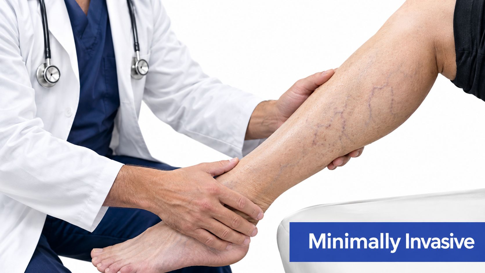
Maria wanted relief from aching, bulging veins, but feared the operation her mother had undergone years earlier. Her specialist explained that radiofrequency ablation uses a thin catheter and ultrasound guidance to close the faulty vein rather than remove it through a larger surgical incision. That distinction often makes RFA feel like a more manageable option, although the treatment still requires a proper diagnosis and recovery plan.
The RFA vein procedure, or radiofrequency ablation, treats venous reflux. Reflux develops when valves in a superficial vein no longer keep blood moving efficiently toward the heart. Blood then travels backward and pools in the leg, contributing to pressure, swelling, aching, skin changes, and varicose veins.
The saphenous vein is one of the main superficial veins in the leg. A refluxing segment can feed pressure into smaller surface veins, so the visible varicosities may be the result rather than the starting point. Occlusion means the treated vein has closed. Blood is redirected through other veins that can carry it effectively.
Why it replaced much of traditional stripping
RFA became an established minimally invasive alternative to surgical vein stripping during the early 2000s. NICE guidance issued in 2003 found that available evidence supported RFA as an alternative to saphenofemoral ligation and stripping when clinicians followed appropriate consent, audit, and governance procedures. The historical recommendation appears in the NICE overview of radiofrequency treatment for varicose veins.
The decision is more involved when a patient also has a wound, diabetes, arterial disease, lymphedema, or leg swelling with several possible causes. Closing a refluxing vein may address one circulation problem without explaining every symptom or improving wound healing on its own. A specialist should therefore review the duplex ultrasound, skin and wound condition, arterial circulation, swelling pattern, and general health before choosing treatment.
The procedure starts with duplex ultrasound mapping. The clinician confirms the direction of blood flow, identifies the refluxing segment, and selects a safe access point. After local numbing medicine, the catheter enters through a small skin opening. The vein stays inside the body, closes, and circulation follows healthier pathways.
Practical rule: Fewer symptoms do not prove that the vein has closed. Duplex ultrasound provides objective confirmation.
How Radiofrequency Energy Closes Varicose Veins
Think of a refluxing vein as a soft, flexible tube with faulty one-way doors. If the doors don't close, blood slips backward whenever gravity works against the leg. RFA doesn't repair those doors. Instead, it removes the malfunctioning pathway from active circulation by shrinking and sealing the vein.
The treatment in clear steps
First, the clinician uses ultrasound to locate the target vein and guide a needle into it. A slender catheter passes through that access point. The team then injects tumescent anesthesia around the vein. This fluid numbs the area, separates the vein from nearby tissue, and helps protect surrounding structures from heat.
Radiofrequency energy travels through the catheter in controlled treatment segments. The energy warms the vein wall rather than heating the entire leg. Heat causes the wall's collagen fibers to contract and the vein to narrow, much like a rubber tube that tightens when its material is gently heated.
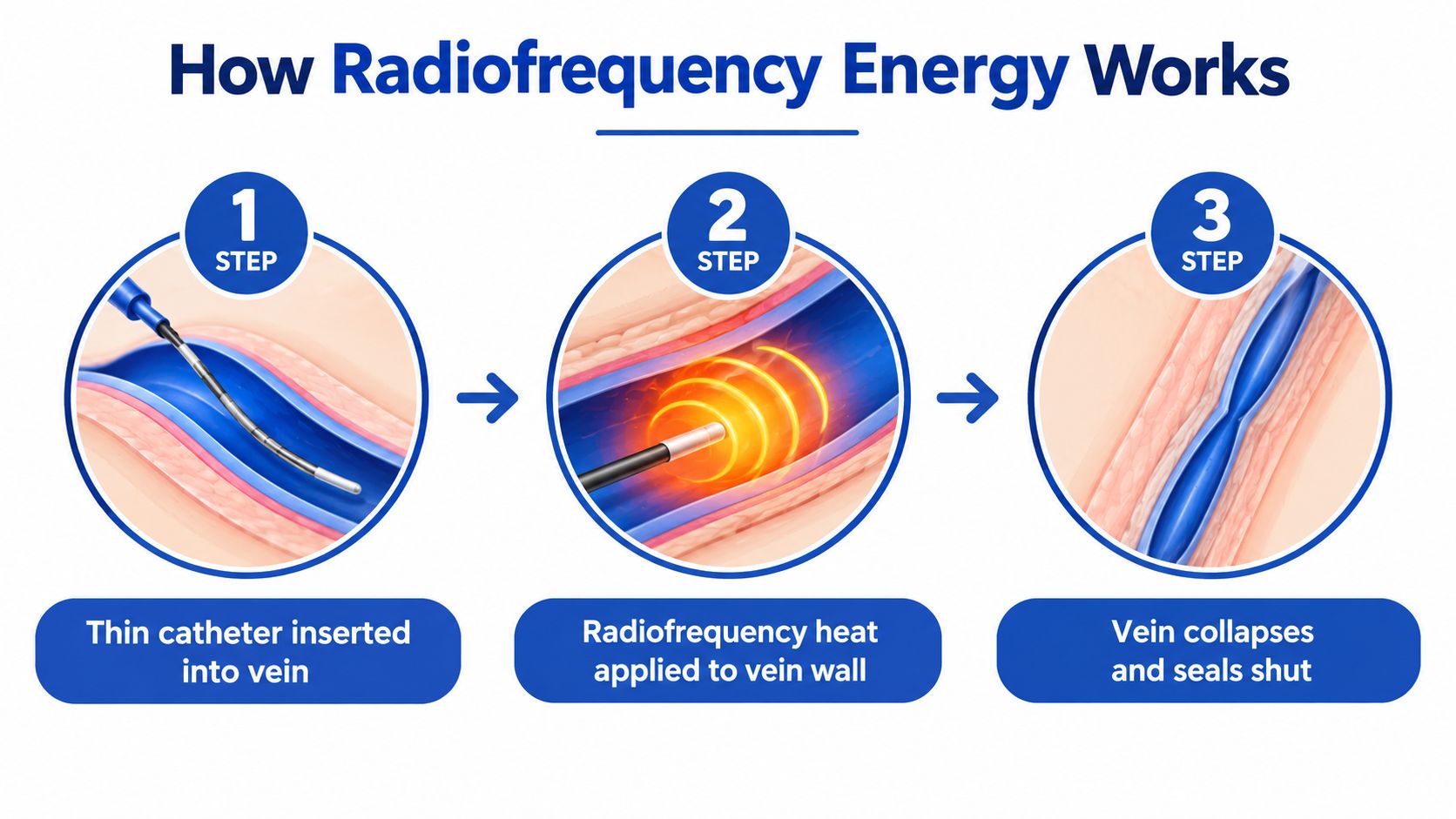
The catheter is gradually withdrawn while the clinician applies energy to the planned sections. Once the treatment is complete, the catheter comes out and the small access point receives a dressing. Blood then travels through healthier superficial and deep veins instead of continuing through the refluxing segment.
What happens after the heat is applied
The immediate effect is contraction and collapse of the vein wall. Over time, the body replaces the closed channel with fibrotic tissue. This is why the treated vein can feel firm or cord-like beneath the skin for a period during healing. The vein's closure, not its removal, is the central result.
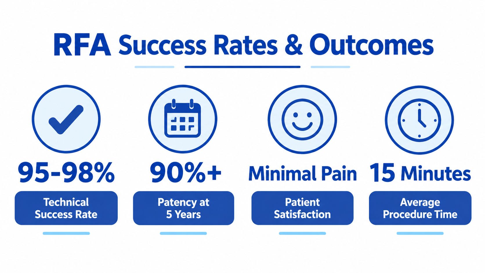
NICE's evidence review reported immediate closure of approximately 90% to 100% of treated long saphenous veins, with reported closure of about 90% to 98% through a maximum follow-up of two years. The same review found that fewer than 5% of patients in the studies continued to report symptoms such as leg pain, fatigue, swelling, or visible varicosities after treatment. These figures come from the NICE evidence overview of radiofrequency ablation, and they describe study outcomes rather than a promise for every individual.
Ultrasound remains important because a vein can reopen, or another vein can later become refluxing. The body's healing response and your symptoms don't always move at exactly the same pace. A technically closed vein may still leave you with swelling if lymphatic, arterial, musculoskeletal, or other venous problems are also present.
What Success Rates and Long-Term Outcomes Look Like
Patients often ask, “What's the success rate?” The most useful answer starts by clarifying what success means. A clinician may be measuring whether the vein is closed, whether reflux has stopped, whether symptoms have improved, whether visible veins have reduced, or whether you need another treatment. Those outcomes overlap, but they aren't interchangeable.
Closure is not the same as permanent symptom relief
A randomized trial with three-year follow-up reported limb-level success of 93.4% after RFA and 88.8% after complete saphenous vein stripping. The difference wasn't statistically significant. In that study, overall treatment failure was 5.2%, including recanalization of 2.9% in the RFA group and surgical failures of 9.4% in the stripping group. Clinical recurrence occurred in 2 RFA patients, or 8.7%, and 3 stripping patients, or 15.8%. These results are summarized in the long-term comparative evidence review.
Recanalization means that a treated vein partially or fully opens again and allows blood flow. Clinical recurrence means that varicose veins or related findings return. A new refluxing vein can cause recurrence even when the original treated vein remains closed. Persistent symptoms may also reflect another untreated source rather than failure of the ablation itself.
A systematic review summarized in Circulation estimated three-year pooled success at 84% for RFA, compared with 94% for endovenous laser ablation and 78% for surgical stripping. Those pooled figures should be interpreted carefully because studies may use different definitions of success, follow patients for different periods, and include different vein anatomies.
Why anatomy changes the outlook
Vein diameter, the length and pattern of reflux, catheter positioning, tumescent anesthesia, device technique, and follow-up definitions can all influence results. A large vein isn't automatically unsuitable for RFA, but it may require careful planning and consistent energy delivery around the entire circumference of the vein.
A meta-analysis of veins larger than 12 millimeters found pooled occlusion of 95.9% and technical success of 99.9%. It also found pooled deep-vein thrombosis of 0.04% and endothermal heat-induced thrombosis of 1.6%. The researchers identified an inverse relationship between increasing vein diameter and occlusion, meaning closure may become more technically demanding as the lumen grows. Those data are reported in the systematic review of thermal ablation in large saphenous veins.
The practical promise of RFA is durable treatment of a documented refluxing segment, not permanent immunity from every future vein problem. Ask which outcome your clinician is tracking, how follow-up will be performed, and whether smaller branch veins may need separate treatment.
RFA Compared with Surgical Stripping and Laser Treatment
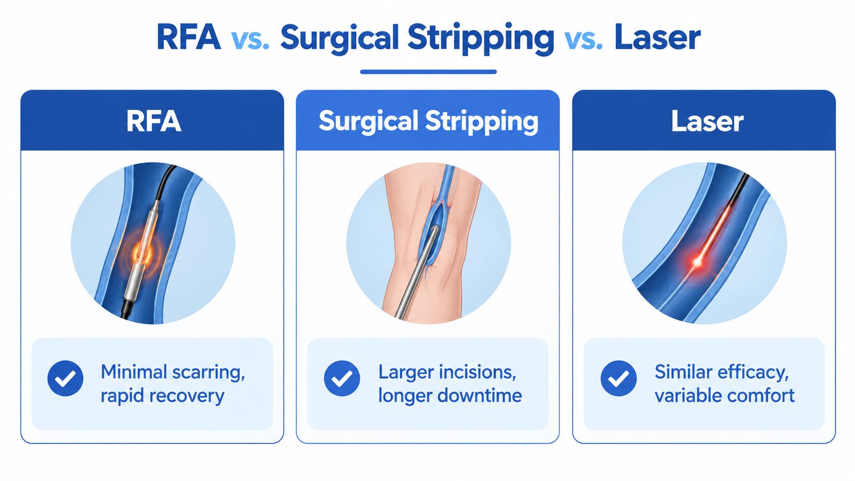
A patient who has lived with aching, bulging veins may welcome a treatment that does not require the incisions associated with surgery. Choosing among RFA, surgical stripping, and endovenous laser ablation still requires more than picking the least invasive-sounding option. Each method closes faulty superficial veins differently, and the best fit depends on vein anatomy, circulation, skin condition, and the recovery demands you can manage.
How the approaches differ
| Treatment | What happens | Practical trade-off |
|---|---|---|
| RFA | A catheter delivers controlled radiofrequency energy inside the vein. | A small access point, no surgical groin incision, and generally faster functional recovery. |
| Surgical stripping | The vein is tied off and removed through surgical access points. | Larger incisions and a recovery that may be more demanding. |
| Endovenous laser ablation | A catheter delivers laser energy to heat and close the vein. | A similar minimally invasive purpose, with comfort and technique influenced by anatomy and equipment. |
A randomized trial compared RFA with laser ablation, ultrasound-guided foam sclerotherapy, and surgical stripping for great saphenous vein incompetence. RFA, laser, and stripping produced comparable vein closure outcomes in the reported follow-up, while foam had a higher rate of persistent reflux. The trial also reported faster recovery and less postoperative pain with RFA than with laser ablation and stripping. Superficial phlebitis occurred more often in the RFA and foam groups.
These findings describe one treatment setting, not a guarantee for every leg. Closure of the treated segment can be successful while another branch, untreated segment, or newly affected vein later causes symptoms. A reasonable expectation for RFA is durable treatment of a documented refluxing segment, not permanent protection from every future vein problem.
What may influence the choice
Laser and RFA both use heat, much like applying controlled warmth to seal a faulty tube, but their energy delivery and treatment technique differ. Vein diameter, tortuosity, proximity to nerves or skin, previous procedures, and the reflux pattern on duplex ultrasound may influence the recommendation. A large or unusually shaped vein may require a method that gives the operator better control.
Patients considering laser treatment can review laser ablation for varicose veins before discussing options with a qualified clinician. The useful question is whether the proposed method can safely treat the refluxing segment while accounting for circulation, skin health, wounds, diabetes, leg swelling, and the expected recovery.
Modern thermal treatment is generally well tolerated, but no procedure is risk-free. Reported major complications include deep-vein thrombosis, pulmonary embolism, and significant nerve injury, while pain is the most commonly reported adverse effect. Careful catheter positioning and tumescent anesthesia matter, particularly when the vein is large or lies close to sensitive structures.
When Vein Treatment Must Include Wound and Circulation Planning
Closing a refluxing vein can be an important part of care, but it isn't a universal cure for every open sore or swollen leg. A patient with a venous ulcer may have superficial reflux, yet healing can remain slow if arterial blood flow is poor, infection is active, compression isn't safe or consistent, or pressure and diabetes continue to damage the tissue.
The first question is not simply whether reflux exists
A duplex ultrasound can show superficial reflux, but the clinician must decide whether that reflux is a meaningful barrier to healing. The evaluation may also need to consider deep-vein patency, arterial perfusion, edema, lymphedema, wound depth, infection, skin viability, mobility, and the patient's ability to use compression.
For example, a leg ulcer with clear venous reflux may benefit from treatment of the refluxing vein, but the wound still requires local management. That can include debridement, appropriate dressings, infection control, pressure reduction, and a compression plan that matches the patient's arterial circulation.
RFA can support a wound-healing plan. It shouldn't replace a wound-healing plan.
The assessment of chronic venous insufficiency should therefore connect symptoms and skin findings with objective circulation testing. This is particularly important for people with diabetes, because neuropathy may reduce pain as a warning signal and diabetes-related changes can complicate tissue repair.
Situations that require coordination
- Venous leg ulcer: Treating reflux may reduce venous pressure, but compression and wound care remain central after ablation.
- Arterial disease: Poor arterial inflow can make compression or intervention unsafe unless circulation is assessed first.
- Lymphedema: Closing a refluxing vein may not resolve lymphatic fluid accumulation, so swelling may need separate treatment.
- Diabetes: Glucose management, foot protection, infection assessment, and regular skin checks can affect healing.
- Active infection: Infection control takes priority when cellulitis, drainage, or systemic symptoms are present.
- Mixed swelling: Heart, kidney, medication, mobility, and musculoskeletal factors can contribute alongside venous disease.
Recent ulcer-focused evidence described 83% freedom from ulcer recurrence at one year after healing, as reported in the 2025 registry report. That finding supports treating venous disease in suitable patients, but it doesn't establish RFA as a standalone wound cure. Your plan should identify what the ablation is expected to improve, what the wound team will manage, and how follow-up will detect stalled healing.
Recovery Timeline and What to Expect After Treatment
Recovery tends to feel more manageable when you know which sensations are expected. The treated vein can feel tight, tender, bruised, or firm as it closes. Mild numbness or tingling can also occur, especially near areas where small sensory nerves run close to the treated vein.
Immediately after the procedure
Your clinician will give individualized instructions about walking, compression, bathing, medication, and exercise. Walking soon after treatment is commonly encouraged because gentle movement supports circulation and prevents long periods of immobility. You may be asked to wear a compression stocking or bandage for a specified period.
Plan a lighter schedule for the rest of the day. Many patients can resume ordinary activities relatively soon, but strenuous exercise, heavy lifting, long periods of sitting, and extended travel may need temporary limits. Don't use a general timeline as permission to ignore your own clinician's instructions.
During the first days and weeks
Bruising may spread slightly before it fades. A pulling sensation, tenderness, redness, or a firm cord along the treated vein can reflect the normal inflammatory response to closure. A small area of numbness may take longer to resolve than the initial soreness.
A 2025 comparative study found 12-month recanalization of 0.40%, symptomatic recurrence of 0.17%, reintervention of 2.62%, and two DVT cases in the RFA group. Another 2025 study reported 96.3% occlusion at three months, while observing pain in 37%, bruising in 14.8%, paresthesia in 7.4%, and thrombophlebitis in 3.7%. These findings are reported in the published 2025 clinical evidence. They help describe patterns in study populations, but they can't predict exactly how your leg will feel.
Contact your clinic promptly if pain is severe or worsening, swelling suddenly increases, redness spreads, the leg becomes unusually warm, you develop significant shortness of breath, or you experience chest pain. A painful, swollen calf also warrants urgent medical advice because deep-vein thrombosis must be assessed rather than dismissed as routine recovery.
Follow-up has a purpose
A scheduled duplex ultrasound checks whether the treated vein remains closed and whether there's any thrombotic extension that needs attention. Follow-up also gives your clinician a chance to examine persistent swelling, a hard tender area, ongoing numbness, skin changes, or varicose veins that may need an adjunctive procedure.
For a patient-friendly preparation guide, review what to expect from a vein procedure. Keep walking as instructed, use compression as directed, and report changes instead of trying to diagnose them from appearance alone.
How to Decide If This Minimally Invasive Option Fits Your Needs
RFA may fit when duplex ultrasound confirms superficial venous reflux, the target vein is accessible, and your symptoms or skin changes match the circulation findings. It may require modification, additional treatment, or a different priority when you have arterial disease, an active wound, significant lymphedema, infection, or unexplained swelling.
Bring these questions to your consultation:
- Which vein is refluxing, and how extensive is the reflux?
- Is the deep venous system open and functioning adequately?
- What improvement should I expect in pain, heaviness, swelling, or skin changes?
- Will visible branch veins need foam treatment or phlebectomy later?
- How will diabetes, arterial disease, a wound, or lymphedema change the plan?
- When will follow-up duplex imaging occur?
- Which recovery symptoms should prompt an urgent call?
The strongest plan connects diagnosis, treatment, wound or circulation management, and follow-up rather than treating the ultrasound image in isolation. If your symptoms are affecting daily life, schedule a vascular evaluation and bring a complete list of medications, medical conditions, prior vein treatments, and wound concerns.
Vein and Wound Institute provides duplex-based vein evaluations, radiofrequency ablation, other minimally invasive vein treatments, and coordinated care for wounds, diabetes-related ulcers, lymphedema, and complex leg swelling. Visit Vein and Wound Institute to request an evaluation and discuss whether RFA belongs in a broader circulation and recovery plan.