
Pitting edema leaves a lasting dent after firm pressure, while nonpitting edema stays firm and leaves no dent. If that distinction is so simple, why can the same person's swelling change from one pattern to the other?
Because the dent describes how the tissue behaves, not what disease someone has. Understanding pitting vs nonpitting edema means reading that observation alongside when swelling started, whether it affects one leg or both, how the skin looks, and whether circulation is adequate.
Table of Contents
- Pitting and Nonpitting Edema Side by Side
- How to Do the Pressure Test and Grade the Pit
- Causes That Produce Each Type of Swelling
- Exam Clues Beyond the Indentation
- Workup, Red Flags and When to Refer
- Management Approaches for Each Pattern
Pitting and Nonpitting Edema Side by Side
The pressure response is one useful clue, but it makes sense only alongside the swelling's distribution and skin changes. Compare the features below rather than treating the pit alone as an answer.
Pitting vs nonpitting edema at a glance
| Feature | Pitting edema | Nonpitting edema |
|---|---|---|
| Pressure response | A temporary indentation remains after pressure | No lasting indentation remains |
| Skin feel | Often soft, with relatively mobile fluid | May feel firm, thickened, or fibrotic |
| Distribution clue | Venous swelling often affects both legs and may vary with standing and elevation | Lymphatic swelling may involve the toes and top of the foot |
These patterns are common associations, not rules that identify a disease. For example, swelling in both legs that changes with position gives different context to a pit than sudden swelling in one leg. Thickened skin or swelling over the toes also changes how a clinician interprets the pressure response.
The pit is a screening observation, not a diagnosis. Read it with laterality, onset, skin changes, and vascular status. A single finding can point toward the wrong workup if it is treated as decisive.
A painful, red, newly swollen leg needs prompt medical review. For other swelling, record which areas are affected and how the skin looks, alongside the pressure response. That description gives a clinician more useful context than the label “pitting” or “nonpitting” alone.
How to Do the Pressure Test and Grade the Pit
The pressure test works by temporarily moving relatively mobile fluid away from the pressed area. Testing over a bony surface makes the resulting indentation easier to see than pressing into a thick layer of soft tissue.
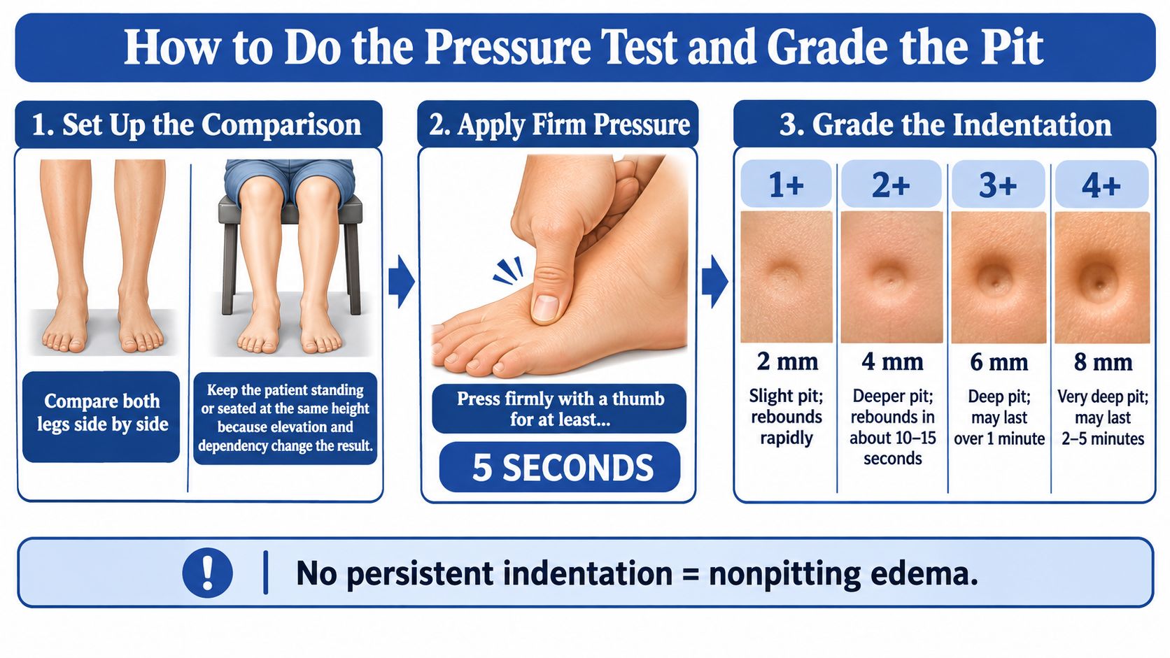
This illustration shows the test setup and grading sequence.
Make the comparison consistent
Position both legs similarly. Sit or stand with the limbs at the same relative height so gravity affects them similarly. Don't compare a leg that has been raised with one that has just been hanging down.
Choose an exposed bony site. Use the shin or the inner ankle over the medial malleolus, avoiding broken skin and painful areas. A sock seam can leave its own mark, so don't mistake clothing impressions for a pressure-test result.
Apply firm, sustained thumb pressure. Hold steadily rather than tapping or rubbing, and use comparable pressure at the matching site on the other leg. Pressing too briefly can make soft, fluid-filled swelling appear nonpitting.
Release, observe, and document. Watch whether a depression forms, estimate its depth, and time the return toward the surrounding skin level. Don't rely on memory when comparing the legs or a later examination.
Use the grade as a description
The commonly used scale summarized in the National Cancer Institute's lymphedema guidance describes 1+ as up to 2 mm with immediate disappearance, 2+ as 2 to 4 mm resolving within 10 to 15 seconds, 3+ as 4 to 6 mm potentially persisting more than 1 minute, and 4+ as deeper than 6 mm potentially remaining for 2 to 5 minutes.
Pitting grade, depth and rebound time
| Grade | Indentation depth | Rebound time |
|---|---|---|
| 1+ | Up to 2 mm | Disappears immediately |
| 2+ | 2 to 4 mm | Resolves within 10 to 15 seconds |
| 3+ | 4 to 6 mm | May persist more than 1 minute |
| 4+ | Deeper than 6 mm | Can remain 2 to 5 minutes |
| Nonpitting | No lasting depression | Tissue stays firm, no rebound dent |
A shallow pit that disappears immediately can still count as pitting. That's different from firm tissue in which no meaningful depression forms.
The scale has limited reproducibility between examiners. Thumb pressure, examination site, and interpretation can differ, so a grade alone isn't a reliable measure of the underlying disease's seriousness.
For a useful chart entry, include:
- Site and technique: Where you pressed and how long you held pressure.
- Response: Estimated grade, indentation depth, and observed rebound.
- Distribution: Which limb was affected and whether the matching site looked similar.
- Skin findings: Softness, firmness, thickening, discoloration, tenderness, or breakdown.
Causes That Produce Each Type of Swelling
The same indentation can have different causes. A pit is a screening clue: its meaning depends on whether swelling affects one leg or both, how quickly it began, and what other symptoms or skin changes accompany it.
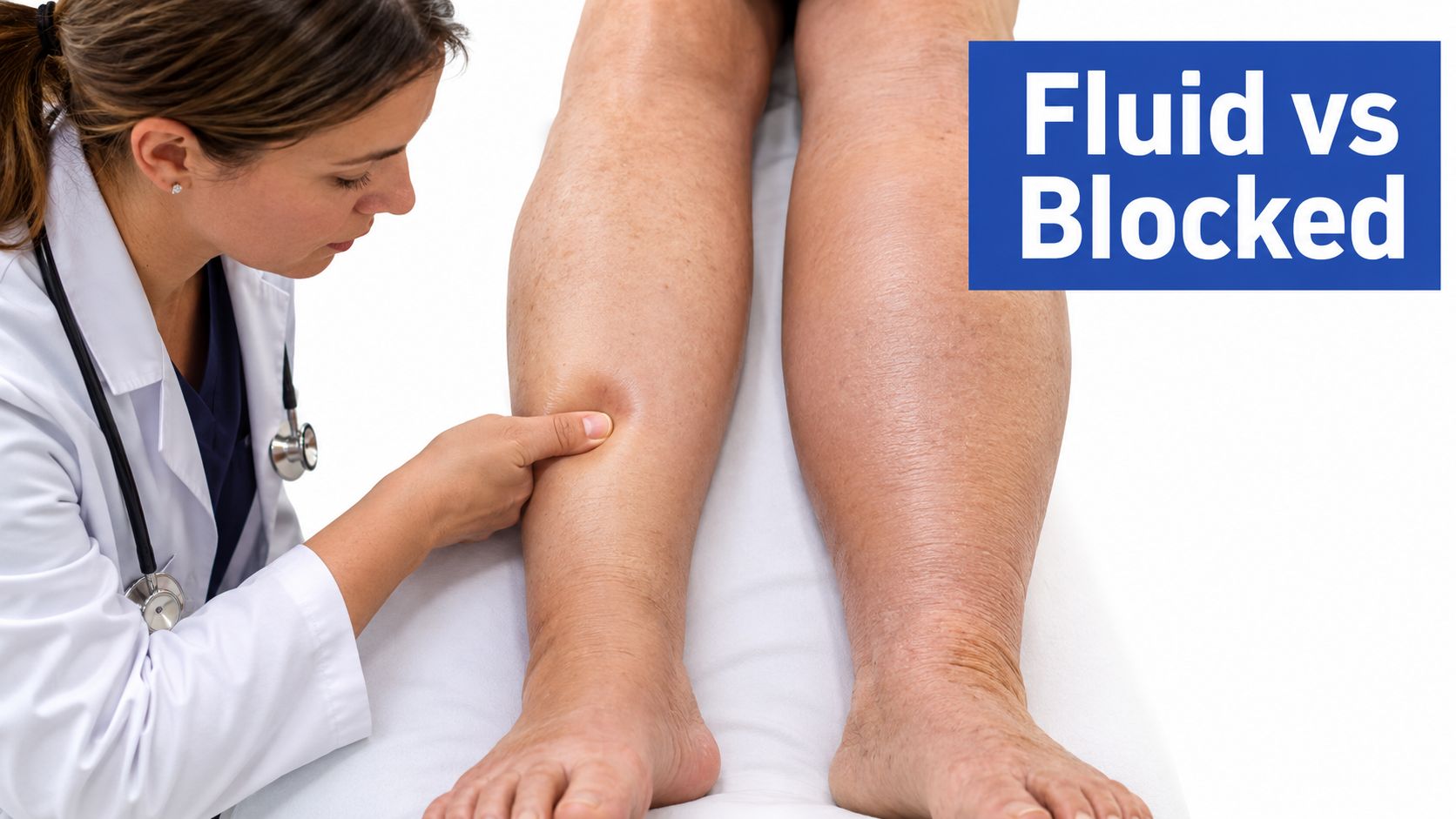
Fluid may collect when pressure in blood vessels pushes it into surrounding tissue, as can happen with chronic venous insufficiency, heart failure, or a deep-vein thrombosis (DVT). Low blood protein, including in nephrotic kidney disease or cirrhosis, can also contribute. Early lymphedema may pit while fluid is still mobile; with longstanding lymphatic dysfunction, tissue changes can make the limb firmer. Hypothyroid myxedema and lipedema are other causes of swelling that may feel firm.
Causes of edema by usual pattern and clue
| Cause | Usual pattern | Clinical clue |
|---|---|---|
| Chronic venous insufficiency | Pitting, often bilateral | Worse with standing, better with elevation, ankle skin changes |
| Deep-vein thrombosis | Pitting, often one-sided | New pain, warmth, redness, or sudden swelling |
| Heart failure | Pitting, often bilateral | Breathlessness, increasing weight, or more widespread swelling |
| Nephrotic kidney disease | Pitting, often generalized | Puffiness around the eyes or protein in urine |
| Cirrhosis | Pitting, often at the ankles | Abdominal fluid or a history of liver disease |
| Lymphedema | May pit early; can become firm over time | Persistent swelling with tissue changes |
| Hypothyroid myxedema | Often nonpitting | Swelling may involve the shins |
| Lipedema | Usually symmetric and nonpitting, with feet spared | Tender fatty tissue and easy bruising |
Consider a person with pitting at both ankles who develops new pain and swelling in one calf. The bilateral ankle pattern may fit a chronic or systemic cause, but the new one-sided change raises a separate concern for DVT. The pit alone cannot settle the question; the new symptom and its timing change what needs to be assessed.
Lymphedema can change in texture as it persists. In a melanoma patient group described in the Vascular Medicine review, 48 patients, or 44%, had stage I lymphedema characterized by pitting, while 61 patients, or 56%, had stage II or III disease. Those figures describe that group, not an individual's likelihood of having lymphedema. They illustrate why a firm or pitting feel alone cannot identify the cause. Learn more about lymphedema and persistent lymphatic swelling.
Causes can coexist. Venous disease and heart failure, for example, may both contribute to ankle swelling. A clinician considers the whole pattern, including whether one or both legs are affected, when swelling began, and whether it changes with position.
Exam Clues Beyond the Indentation
A useful examination connects tissue response with the foot, skin, veins, symptoms, and limb size. Each clue changes which explanation is more plausible, but each also has limits.
Read the skin and swelling distribution
Stemmer sign: The lymphoedema best-practice document describes a positive sign as inability to pinch and lift a skin fold at the base of the second toe or finger, and notes that approximately 10% difference between affected and unaffected limb measurements is used by many practitioners to support diagnosis. A positive sign strongly supports lymphatic disease, but a negative sign doesn't exclude an earlier or more proximal presentation.
Skin texture: Soft tissue suggests a more fluid-dominant component. Thickened, rubbery, warty, or hyperkeratotic skin, meaning an abnormally thick outer skin layer, supports chronic tissue change but doesn't reveal every contributing cause.
Toe and foot involvement: Swelling over the top of the foot and into the toes strengthens suspicion for a lymphatic pattern. Foot sparing can help distinguish another pattern, such as lipedema, but footwear and body shape can make the boundary difficult to judge.
Venous changes: Varicose veins, brown ankle pigmentation, hardening near the ankle, and ulceration support venous assessment. Visible veins alone don't establish why the entire limb is swollen.
Warmth and tenderness: These findings change the urgency, especially if they're new. They shouldn't be dismissed as routine consequences of chronic swelling.
Measure what can change
Use a flexible measuring tape at reproducible anatomical landmarks, with consistent limb position and tape tension. Record the landmarks so the next examiner can measure the same sites rather than choosing a different part of the calf.
Circumference and limb-volume tracking can be more actionable than repeatedly assigning a pit grade. However, a comparison with the opposite limb is less informative when both limbs are affected, and normal asymmetry must be considered.
Document onset, laterality, positional changes, and current medications. Bring a medication list rather than stopping a suspected medicine yourself, because the prescribing clinician needs to weigh its benefits against the swelling.
A better follow-up question is whether the limb is becoming smaller, softer, healthier, and more functional, not simply whether it still pits.
Pitting with venous signs supports venous evaluation, with arterial status assessed before choosing compression. Firm nonpitting swelling with a positive Stemmer sign supports lymphatic evaluation, skin-care counseling, infection prevention, and measurement-based monitoring.
Workup, Red Flags and When to Refer
A pit is a screening clue, not a diagnosis. The workup depends on what else is happening: whether swelling is new or longstanding, affects one or both legs, and comes with pain, skin changes, or signs of vascular or systemic illness. A test should answer a specific clinical question.
| Test | What it checks | When it is ordered |
|---|---|---|
| Venous duplex ultrasound | Reflux, venous obstruction, or deep-vein thrombosis | Unilateral swelling, venous skin changes, new pain, or suspected chronic venous disease |
| Arterial pressure testing | Adequacy of arterial blood supply | Before selecting compression, especially with wounds, ulcers, or rest pain |
| Kidney function and urine protein | Kidney-related fluid retention or protein loss | Generalized or bilateral swelling with suggestive symptoms |
| Albumin and liver panel | Low blood protein and liver-related causes | Generalized swelling, abdominal fluid, or liver history |
| Thyroid-stimulating hormone | Thyroid dysfunction | Firm nonpitting swelling with dry skin, fatigue, or other thyroid symptoms |
| Echocardiogram or natriuretic peptide testing | Heart function and possible cardiac fluid overload | Bilateral swelling with breathlessness or increasing weight |
| Lymphoscintigraphy | Lymphatic transport | Uncertain lymphedema, generally in specialist settings |
This is a menu of clinical questions, not a checklist for every person with edema. Examination and medical history guide which tests, if any, are appropriate.
Let urgency determine the first step
A newly painful swollen leg raises an immediate question: could a clot or infection need treatment? Venous duplex ultrasound is commonly prioritized when thrombosis is suspected. Assessing chronic venous reflux may require a different ultrasound protocol.
The BC Cancer lymphedema guidance describes applying firm pressure for at least 5 seconds and interpreting the result alongside the broader examination. New one-sided pitting with redness, warmth, tenderness, or acute pain needs particular attention.
Generalized swelling may call for a different set of questions. Kidney function, urine protein, albumin, and liver tests can help assess fluid retention, protein loss, or reduced protein production. Breathlessness may prompt cardiac testing; firm swelling with thyroid symptoms may justify thyroid-function assessment.
Arterial testing and venous ultrasound answer different questions. Venous reflux does not show whether the arteries deliver enough blood for a particular compression plan.
Symptoms that override a reassuring-looking pit
Seek same-day medical assessment for:
- New one-sided swelling with pain, redness, warmth, or tenderness: A clinician needs to assess for thrombosis, cellulitis, and other acute causes.
- Sudden swelling after surgery, prolonged travel, or immobility: Explain the timing and circumstances when arranging care.
- Rapid progression: A limb becoming noticeably larger or tighter needs reassessment, even if swelling was previously diagnosed.
- Fever or a concerning wound: New drainage, odor, spreading redness, or worsening wound pain warrants prompt review.
- Swelling with increasing breathlessness or rapid weight gain: These symptoms can require urgent systemic evaluation.
Chest pain, severe breathlessness, fainting, or coughing up blood with leg swelling require emergency care. Do not wait for a routine vein appointment or try to resolve these symptoms with elevation.
A painful one-sided pitting leg should not be managed with compression alone while thrombosis or cellulitis remains unassessed. The absence of a pit does not rule out a serious cause.
Match the referral to the unanswered question
Stable swelling in both ankles that becomes more noticeable by evening can start with a primary-care assessment. Reviewing medications and considering venous, cardiac, kidney, or liver contributors helps prevent visible varicose veins from being mistaken for the whole explanation.
Persistent swelling with heaviness, ankle discoloration, or a wound in a venous pattern may warrant vascular assessment. The guide to when to see a vein specialist describes symptoms that may merit that discussion. An evaluation can help determine the severity of chronic venous disease.
Firm swelling involving the toes, substantial skin thickening, or persistent suspected lymphatic dysfunction supports referral for lymphedema assessment. If the diagnosis remains uncertain, a specialist can determine whether lymphatic imaging would affect the next steps.
For a closer look at how clinical reasoning works, consider how clinicians weigh multiple clues rather than relying on one finding. The practical question is which explanation fits the full presentation and which dangerous alternative must be excluded.
Vein and Wound Institute treats vein conditions and chronic wounds using minimally invasive techniques. Its wound services include care for diabetic, venous, arterial, pressure, post-surgical, traumatic, and non-healing wounds, as well as wound infections.
A useful workup produces findings that can change the plan. A duplex showing thrombosis calls for a different response from uncomplicated reflux, while low albumin directs attention to why blood protein is reduced.
Management Approaches for Each Pattern
| Assessed cause or pattern | Management approach | When to seek reassessment |
|---|---|---|
| Venous pitting edema | Elevation, movement and activity changes, plus graduated compression when vascular assessment supports it | Skin breakdown, an ulcer, worsening symptoms, or insufficient improvement |
| Lymphedema with firm or nonpitting tissue | Individualized decongestive therapy, potentially including manual lymphatic drainage, bandaging or garments, exercise, and skin hygiene | Redness, fever, rapid enlargement, or skin breakdown |
| Heart-, kidney-, or liver-related edema | Treatment of the underlying disease, with prescribed medication and monitoring | Breathlessness, falling urine output, or increasing abdominal swelling |
| Hypothyroid myxedema | Thyroid treatment guided by testing and clinical review | Persistent or worsening swelling despite treatment |
| Acute deep-vein thrombosis | Prompt clinician-directed treatment, commonly anticoagulation; compression only under an individualized plan | Chest pain, breathlessness, or worsening symptoms |
Compression follows assessment, not the dent
A visible pit is one clue, not a compression prescription. The AMBOSS overview of peripheral edema describes why assessment of vascular and systemic causes matters before treating a swollen leg.
Before recommending compression, a clinician considers arterial circulation, possible infection or thrombosis, the distribution of swelling, and the cause of any wound. Properly selected compression can support venous ulcer healing, but the garment and pressure must suit the person's circulation and skin condition.
Avoid borrowing someone else's stockings or assuming a tighter garment will work better. A clinician can select an appropriate approach. New pain, numbness, or changes in foot color or temperature call for prompt review.
For edema related to chronic venous insufficiency, supportive measures may ease symptoms. Persistent symptoms despite compression, elevation, and activity changes may warrant consideration of a minimally invasive procedure. The choice depends on vein size and location, ultrasound findings, symptoms, and overall vascular health.
Treat the source of the swelling
Lymphatic care may include manual lymphatic drainage, appropriate compression, movement, and skin protection as part of an individualized plan. Painful, unexplained swelling should be assessed before starting treatment.
Diuretics are prescribed for particular conditions, not as a general response to any swollen ankle. Take them as directed, and contact the prescribing clinician rather than changing the dose when swelling continues.
Treatment principle: Choose the plan for the cause and the circulation.
Vein and Wound Institute provides evaluation and treatment for venous conditions, leg swelling, lymphedema, poor circulation, and complex wounds. When swelling and a wound occur together, assessing circulation can help guide care for both.
Vein and Wound Institute offers evaluation of leg swelling, venous disorders, lymphedema, and complex wounds, with onsite diagnostics and coordinated vein and wound care. For persistent swelling without emergency symptoms, visit Vein and Wound Institute to arrange an assessment and learn which findings should guide your next step.