Picking the best laser first is the most common mistake I see people make. A device doesn't decide your result. Vessel type, depth, diameter, and skin type do. Fine red telangiectasias on the cheek, a blue reticular vein at the temple, diffuse rosacea redness, and a larger nasal vessel aren't the same condition, and they shouldn't get the same treatment.
That's how this comparison of facial vein treatment options is organized. Each option below is matched to the vessels it suits, with its real trade-offs in downtime, pigment risk, pain, and recurrence. Some are precise and inexpensive but slow. Others clear broadly but need repeat sessions. A few are excellent for deeper veins and wrong for fine surface ones.
Start by asking what kind of vessel you have. Then choose the tool that fits it.
Table of Contents
- 1. Microsclerotherapy for Facial Spider Veins
- 2. Pulsed Dye Laser (595 nm) for Facial Telangiectasias
- 3. Nd:YAG Laser (1064 nm) for Deeper Facial Veins
- 4. Intense Pulsed Light (IPL) for Facial Redness and Vessels
- 5. Bipolar Electrodesiccation (Electrocautery) for Facial Vessels
- 6. Ultrasound-Guided Foam Sclerotherapy for Larger Facial and Nasal Veins
- 7. Diode Laser (980 nm) for Facial Telangiectasias
- Facial vein treatment options at a glance
- Matching the Treatment to the Vessel, Not Just the Redness
1. Microsclerotherapy for Facial Spider Veins
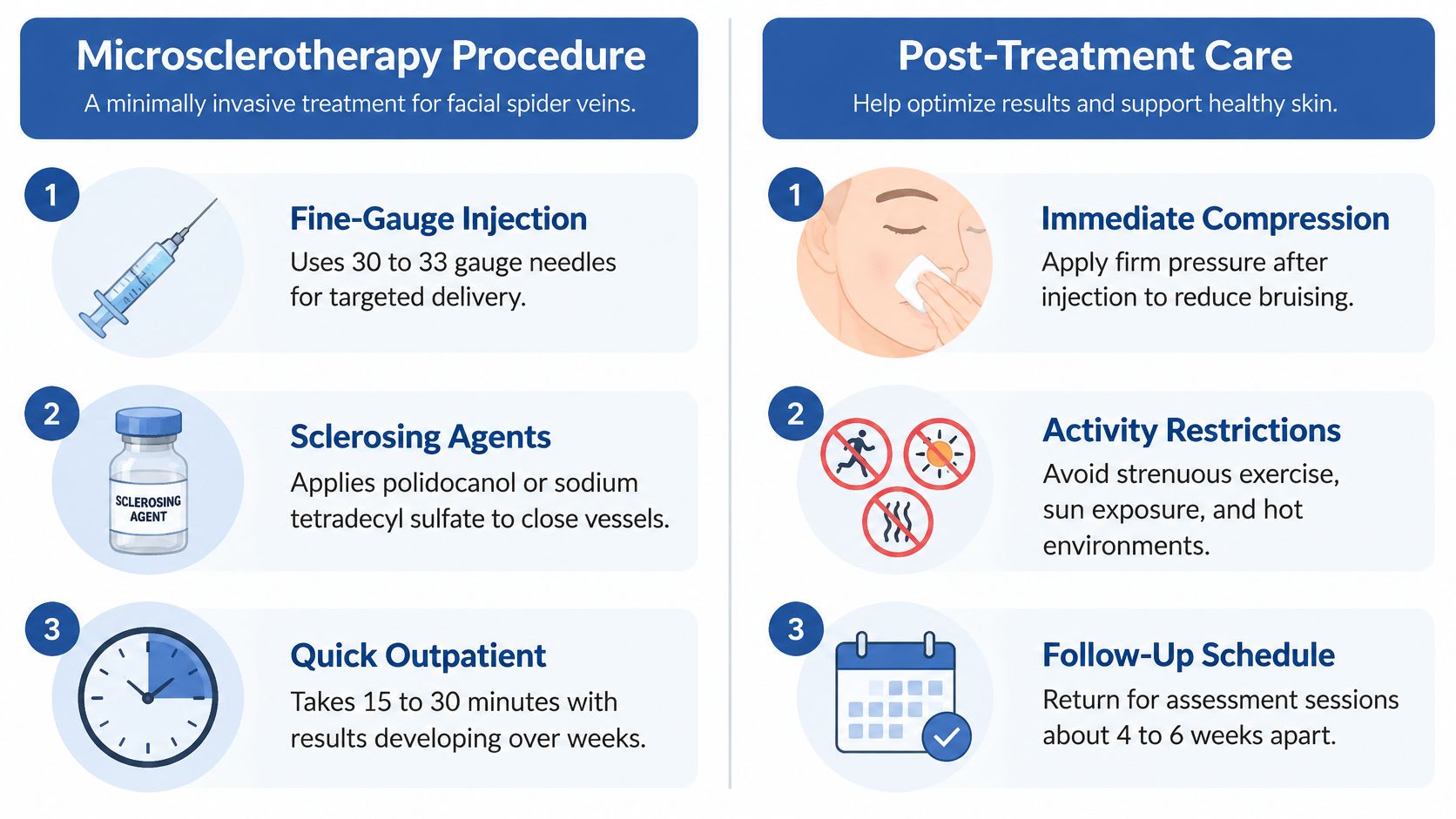
Microsclerotherapy injects a tiny amount of polidocanol or sodium tetradecyl sulfate through a very fine needle directly into a small facial vessel. The agent irritates the vessel wall, the vein closes, and your body gradually absorbs it. For discrete perinasal and malar telangiectasias on the cheeks, nose, and chin, that precision is hard to beat.
It isn't universal. Very small, superficial facial vessels near sensitive structures aren't always suitable for injection, and anatomy should drive the choice. A retrospective review in Dermatologic Surgery of more than 300 patients treated with sclerotherapy reported approximately 50% to 100% resolution, depending on the sclerosant and clinical circumstances. That wide range is the point. Selection matters.
Where It Works Best
Say you have a handful of stubborn red vessels around the nose that remain after laser treatment. A clinician can use microsclerotherapy for those residuals, often alongside pulsed dye laser for partial clearance. If you're weighing injection against a thermal probe approach, read this comparison of liquid sclerotherapy and VeinGogh for facial spider veins before deciding.
- Use the lowest effective concentration: Facial skin shows pigment change easily, so restraint protects the result.
- Apply firm pressure after injection: It limits bruising and helps the vessel stay closed.
- Avoid heat and strenuous exercise for several days: Sun, saunas, and hard workouts dilate vessels you just treated.
- Reassess in 4 to 6 weeks: Don't judge clearance until the vessel has had time to be absorbed.
Expect possible incomplete clearance, transient redness, swelling, or pigment changes, and plan for more than one session if vessels are numerous.
2. Pulsed Dye Laser (595 nm) for Facial Telangiectasias
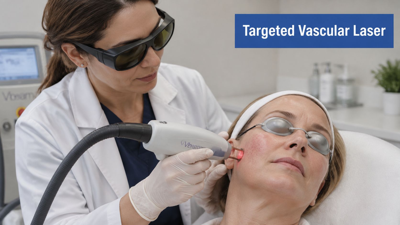
For fine, superficial red vessels, pulsed dye laser at 595 nm is the most vessel specific light option on this list. Hemoglobin absorbs that wavelength strongly, so the energy heats and coagulates the vessel while sparing most surrounding skin. Platforms such as the Candela Vbeam are used this way for facial telangiectasias and for diffuse redness with visible vessels in rosacea.
Depth is its limit. A 2023 study comparing 595-nanometer pulsed-dye laser and 1064-nanometer Nd:YAG laser found that pulsed-dye laser performed better for smaller, more superficial vessels, whereas Nd:YAG suited deeper spider veins. If your vessels are blue, deeper, or larger caliber, pulsed dye alone will often underdeliver.
Getting a Clean Result
Cooling isn't optional. It reduces epidermal injury and discomfort, especially when treating the nose and upper lip where skin is thin. Test a small area first if you have a darker skin type, because melanin competes for the energy and raises pigment risk.
Practical rule: Photograph the area in the same light before every session. Memory flatters or disappoints. Photos don't.
Daily broad-spectrum sun protection before and after treatment protects against post treatment pigmentation. Bruising, swelling, crusting, or blistering can occur, and good aftercare shortens all of them. Follow these steps for caring for your skin after facial vein treatment rather than improvising with active skincare.
I recommend pulsed dye when the problem is fine red vessels and diffuse erythema close to the surface. I don't recommend it as a first move for a prominent blue temple vein.
3. Nd:YAG Laser (1064 nm) for Deeper Facial Veins
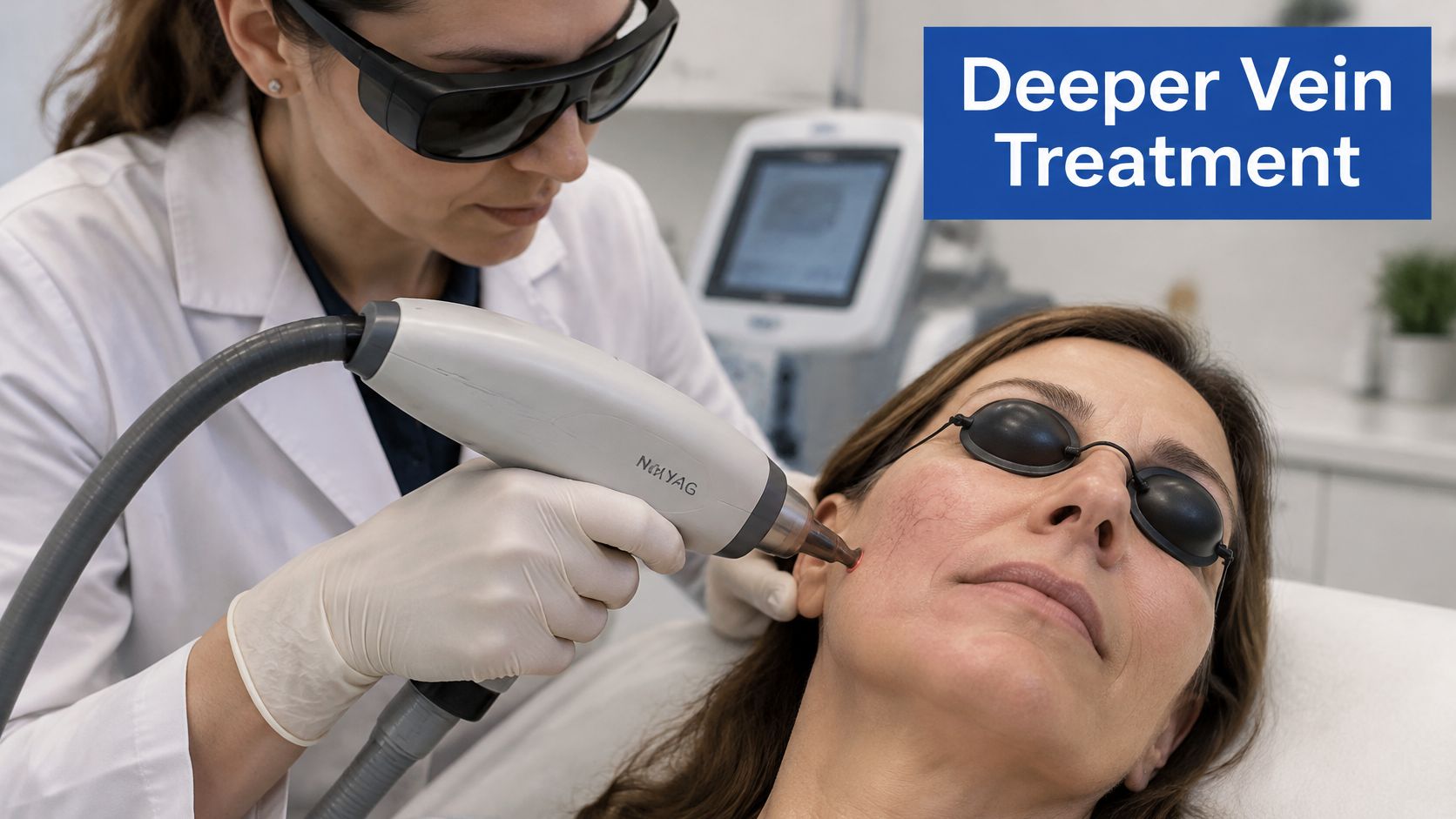
Nd:YAG at 1064 nm penetrates deeper than visible wavelength lasers. That makes it the practical choice for larger or deeper facial veins, including some blue reticular vessels, and its longer wavelength is less strongly absorbed by melanin, which helps in darker skin tones. The blood inside the vessel absorbs the energy, heats, and the vessel closes. A superficial device can't reach that depth, so a deeper component survives the session and reappears within months.
Caliber decides more than device brand. A systematic review of six clinical trials involving 271 patients reported clearance rates of 95–100% for 1064-nm Nd:YAG lasers with generally limited adverse effects, and in its comparative data 72% of patients rated Nd:YAG outcomes as good to excellent at one year compared with 48% for IPL, with the gap widest for vessels larger than 1 mm.
Safety Around the Eyes
Periocular vessels demand strict eye protection for patient and staff, conservative starting energy, and cooling before, during, and after each pulse. When a deeper cheek vessel sits beneath a mat of fine surface redness, treat in sequence rather than all at once: Nd:YAG first for the deeper vessel, then sclerotherapy or a superficial laser for residuals at follow-up in 4 to 8 weeks.
Depth also earns you a diagnostic pause. A facial vein that returns after a technically sound Nd:YAG session, or one that keeps enlarging, deserves investigation rather than another pass. Re-treating is easy. Explaining a recurrence you never looked into is not. Persistent surface vessels can be the visible end of deeper venous reflux, and no laser at this wavelength corrects the source.
Clinics exploring multi application Nd:YAG platforms can discover Fotona 4D facial treatments to see how the wavelength is packaged. The platform name matters less than whether your clinician documents vessel diameter and morphology and titrates settings to your response.
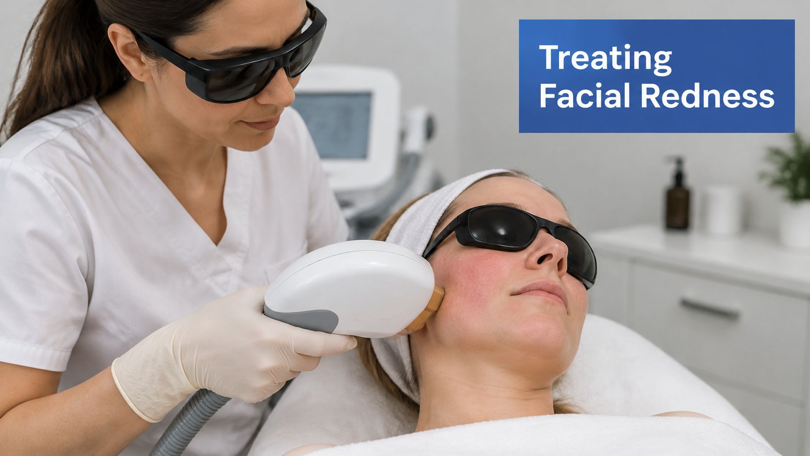
4. Intense Pulsed Light (IPL) for Facial Redness and Vessels
IPL uses broadband, filtered light that targets hemoglobin and melanin across many wavelengths at once. That breadth is the point: rosacea flushing, scattered fine telangiectasias, and a decade of sun damage all improve in the same pass, which a single-wavelength laser cannot manage. It is also the limit. One dilated vein at 1.5 mm, or a vessel fed by a deeper refluxing tributary, will not close reliably when the energy spreads across a band of wavelengths.
Results are real but variable. A systematic review of IPL in rosacea in the Indian Journal of Dermatology, Venereology and Leprology summarized studies reporting improvement in 95.3% of patients at six months, with 66.4% classified as effectively treated. Typical short term effects included erythema, flushing, edema, bruising, and mild burning that generally resolved spontaneously.
Who IPL Suits
The candidate I would book describes an all-over cheek flush that flares with heat, wine, or exercise, plus fine vessels and blotchy pigment, rather than three veins she can point to. IPL covers that picture in one session and pairs naturally with topical rosacea therapy for the underlying inflammation. A 2026 retrospective study in Dermatology and Therapy of 111 patients treated with broadband light for facial vascular lesions found localized microcrusting in 17.1% of patients, with no serious adverse events reported.
- Patch test and set fluence by Fitzpatrick type: tanned or darker skin is a risky mix, because melanin competes with the hemoglobin you are trying to reach.
- Avoid sun before and after: sunscreen is part of the treatment, not an extra.
- Plan for multiple sessions: maintenance is typical, especially with rosacea.
One judgement worth stating plainly: redness that stays on one side, or a cheek vessel that fills when the patient leans forward, deserves a duplex ultrasound before any light session. That pattern can be the facial sign of deeper venous reflux, and IPL will fade the surface while the source keeps feeding it. Understanding triggers helps too, so review what causes facial veins and how to prevent them alongside treatment.
5. Bipolar Electrodesiccation (Electrocautery) for Facial Vessels
Electrodesiccation is the oldest tool here, and it still earns its place. A fine probe delivers a controlled electrical current into a single vessel, closing it through localized heating. No light, no injection, no expensive platform. For one isolated spider vein or a cherry type spot that hasn't responded to laser or sclerotherapy, it's direct and inexpensive.
Precision cuts both ways. You're creating a tiny thermal injury, so technique determines whether you get clearance or a scar. This isn't a tool for widespread redness or a full cheek of fine vessels. Treating dozens of vessels one by one is slow, uncomfortable, and raises cumulative skin injury.
How I Limit Scarring Risk
Use the smallest effective current and probe tip, treat one vessel at a time, and let skin recover between passes rather than chasing every branch in one sitting. Wound care afterward is simple but non negotiable. Keep the spot clean, don't pick the crust, and protect it from sun while it heals.
Reserve electrodesiccation for targeted, stubborn lesions. If a clinician proposes it for diffuse redness across both cheeks, ask why a light based option isn't safer.
A typical use case is a residual vessel at the nostril rim after sclerotherapy. One or two precise applications can finish what injection couldn't reach. Expect a small crust for several days, possible temporary pigment change, and a result you judge only after the skin has fully settled. For the right single lesion, that trade is worth it.
6. Ultrasound-Guided Foam Sclerotherapy for Larger Facial and Nasal Veins
A nasal or temple vein that keeps refilling after surface treatment points to a feeder vessel or a deeper connection. That pattern changes the choice of tool. When a facial vein is larger, deeper, or part of a broader venous network, liquid injection by eye alone is not enough. Foam sclerotherapy turns the sclerosant into microfoam that displaces blood and spreads along the vessel wall. Duplex ultrasound guidance lets the clinician see the vessel and confirm placement before injecting.
The imaging step separates cosmetic touch-up from medical vein care. A review of seven studies involving 771 patients reported an odds ratio of 5.64 for greater efficacy compared with a comparator treatment, efficacy of 75.7% at one year, pigmentation in 14.9%, and residual telangiectasia in about 15% (review of foam sclerotherapy studies). Higher efficacy comes with visible trade-offs on facial skin. Pigment change and residual vessels are real limits, not rare exceptions.
When to Insist on Ultrasound
A prominent nasal or temple vein that persists after surface treatment suggests a feeder or deeper connection. The treatment plan has to be based on the source of the problem, not only on the veins visible at the skin's surface. A consultation with duplex ultrasound helps identify where abnormal reflux is occurring and which treatment is appropriate.
- Confirm anatomy first: Map the vessel and nearby structures before any injection.
- Use extreme care near the eye: Avoid areas with known arterial supply risks.
- Monitor for visual symptoms: Document them and give clear written aftercare.
- Coordinate with a vein specialist: Essential for venous malformations or reflux-driven vessels.
I reserve ultrasound-guided foam for veins that fine surface treatment cannot clear. For superficial telangiectasias, a superficial laser is the lower-risk tool.
7. Diode Laser (980 nm) for Facial Telangiectasias
The 980 nm diode laser sits between pulsed dye and Nd:YAG in the way it behaves on facial skin. Hemoglobin absorbs its wavelength well, melanin absorbs it less than with shorter wavelengths, and the hardware is compact enough to run in an office setting. For targeted facial vessels in a patient whose main worry is pigment change, that balance makes it a reasonable middle option.
What one wavelength can deliver is usually meaningful clearance over several sessions, not erasure in a single visit. The widely cited clearance figures in diode marketing often trace back to Nd:YAG work rather than diode work, so treat cross-device comparisons with caution until you see data on the specific device in front of you. In practice I judge a diode result by how the vessel blanches and clears session by session, not by a quoted percentage borrowed from another laser.
A Worked Example
Consider a typical mixed pattern: fine red vessels across the cheeks plus a few slightly larger ones along the jawline. A clinician may treat the larger vessels with diode laser and reserve a different approach, such as sclerotherapy, for what remains, rather than forcing one device to cover everything. That is sound clinical sequencing, not upselling.
Start with lower energy and titrate upward based on the vessel's immediate response. Cool during and after treatment to reduce discomfort and surface heating. Avoid heat and sun exposure afterward, since both provoke redness in freshly treated skin.
Photograph the area the same way each visit and reassess before deciding on another pass. If a vessel has not changed after two well chosen treatments, switching modality usually beats raising energy on the same device. Facial skin has little tolerance for repeated high-fluence passes, and the discipline of stopping early protects it.
One pattern deserves a referral rather than another laser session. A prominent vessel that refills after surface treatment, or redness that spreads beyond a discrete vessel, can be a surface sign of deeper venous reflux. Our treatment planning starts from the source of the problem, not from what shows at the skin's surface, and duplex ultrasound during a consultation identifies where abnormal reflux is occurring. Lasers treat what they can see. They do not fix what feeds it.
Facial vein treatment options at a glance
Read this table by vessel and skin type, not by device name. A fine cheek vessel and a blue temple vein are different problems even when both look red in a photograph.
| Item | Best for | Typical downtime | Typical sessions | Main risk |
|---|---|---|---|---|
| Microsclerotherapy for Facial Spider Veins | Small, isolated vessels with a clear feeding point | Temporary bruising or redness | Multiple; spaced weeks apart | Pigmentation changes; rare skin injury |
| Pulsed Dye Laser (595 nm) for Facial Telangiectasias | Fine vessels and diffuse redness when purpura is acceptable | Short-lived redness or purpura | Multiple; spaced weeks apart | Temporary purpura or pigmentation changes |
| Nd:YAG Laser (1064 nm) for Deeper Facial Veins | Larger or deeper blue facial veins | Bruising or discomfort may occur | Follow-up, repeat only if needed | Burns or scarring if settings are too high |
| Intense Pulsed Light (IPL) for Facial Redness and Vessels | Diffuse redness, fine vessels, and pigment | Temporary redness or swelling | Multiple; spaced weeks apart | Pigment changes if settings are poorly calibrated |
| Bipolar Electrodesiccation (Electrocautery) for Facial Vessels | Single stubborn spots, not a field of redness | Pinpoint crust; local wound care | Targeted; reassess at follow-up | Scarring or skin texture changes |
| Ultrasound-Guided Foam Sclerotherapy for Larger Facial and Nasal Veins | Larger facial or nasal veins with a visible feeder | Bruising or swelling may occur | Reassess after initial treatment | Foam migration, including possible visual disturbance |
| Diode Laser (980 nm) for Facial Telangiectasias | Small to medium facial vessels | Temporary redness, swelling, or bruising | Multiple; reassess between sessions | Temporary skin reactions or pigment changes |
The choice is not a ranking. Ultrasound-guided foam treats a larger vessel with a feeder, but carries a migration risk that surface lasers do not. Electrodesiccation is precise for one point and a poor fit for a broad field of redness. If a facial vein refills quickly, returns after good treatment, or is fed by a larger branch, it may be a surface sign of deeper venous reflux. Closing the visible branch alone gives a short-lived result; the plan has to follow the source of the problem, not just the veins visible at the skin's surface.
Matching the Treatment to the Vessel, Not Just the Redness
Anatomy determines the option. Vessel size, depth, wall thickness, and the skin overlying it decide whether a surface treatment will work or just irritate the face. Fine isolated vessels behave differently from diffuse redness, and a deeper blue vein under thin skin needs a different wavelength and penetration depth than a nasal telangiectasia. The table above maps those categories to devices. Use it as a starting point and confirm the choice with ultrasound.
Before you book, answer one question: is this vessel purely cosmetic, or is it a surface sign of deeper venous reflux? A temple or under-eye vein that refills quickly, a cluster fed by a larger reticular vein, or vessels that return after good treatment all point to a source below the skin. Closing the visible branch without addressing the feeder gives you a temporary result and a repeat bill. That distinction matters more than the device name, because a facial vein can be the visible tip of a drainage problem that a laser alone will not solve.
Duplex ultrasound answers the question directly. It shows vessel size, depth, flow direction, and connections you cannot see in a mirror. At Vein and Wound Institute, recommendations are personalized based on your condition and goals, and the choice of procedure depends on the size and location of the vein, ultrasound findings, symptoms, and overall vascular health. That assessment turns a device menu into a treatment plan.
Set expectations realistically. Expect repeat sessions spaced weeks apart for most devices, and expect fading over weeks rather than days. New vessels can appear over time with sun exposure, rosacea, and aging. Gentle aftercare protects the result, and if your skin is reactive, it helps to understand caring for sensitive skin with aloe before layering active products back on.
Choose a clinician who measures your vessels, photographs them, explains why a specific option fits your skin type, and tells you what it will not do. That is worth more than any device name. Bring your questions, ask about pigment risk and retreatment rates, and do not book until the plan matches the vessel in front of you.
If you are weighing facial vein treatment and want to know whether your vessels are cosmetic or connected to deeper reflux, Vein and Wound Institute offers vein evaluation with duplex ultrasound and minimally invasive treatment plans. Visit Vein and Wound Institute to request a consultation and get a plan matched to your vessel type, skin, and goals.