
An ankle-brachial index can come back normal while a foot ulcer stays open week after week. That pairing confuses many patients, and it starts to make sense once you see how diabetes and circulation interact. Two failures stack at once. Damaged vessels cut the supply line, and damaged nerves hide the injury, so you may never feel the blister that started it.
The limb works like a city block. Large arteries and tiny capillaries are the pipes. Nerves are the sensors that call for help. Tissue cannot rebuild when the pipes narrow and the alarm never rings.
Because the two failures travel together, they have to be measured together. The useful next step is to match what you feel, and what you do not feel, to the kind of damage in the limb.
Table of Contents
- Diabetes and Circulation at a Glance
- Why Poor Flow Turns a Small Wound Into a Limb Threat
- Blood Flow Tests and the Numbers That Matter
- Telling Arterial, Venous and Nerve Wounds Apart
- What to Do When a Diabetic Wound Stops Healing
- What Treatment Looks Like When Flow Is Low
- Prevention and Coordinated Care That Protects Limbs
- Book a Circulation Check If Warning Signs Appear
Diabetes and Circulation at a Glance
Many patients arrive with one question. Is this a blood-flow problem, a nerve problem, or both? The answer changes which test matters and which care path comes next.
Large-artery narrowing, called peripheral artery disease, sits in the leg and foot arteries. Plaque narrows the pipe. Calf muscles cramp when you walk, the foot feels cold, and pulses may be weak or missing.
Small-vessel damage sits in the capillaries of the skin. Walls thicken, so fine oxygen exchange falls even when a big pulse is still there. Cuts linger, skin thins, and redness hangs on.
Neuropathy is the sensor failure. Numbness or tingling is common. Plenty of people feel nothing at all, so a shoe rub or a thick callus never registers as an injury.
The combined row is the one that threatens the limb. Poor delivery and an unnoticed injury compound each other. The wound is often found late, and infection is already underway.
| Damage route | Where it happens | What it does to blood flow | What you may notice |
|---|---|---|---|
| Large arteries (peripheral artery disease) | Leg and foot arteries | Atherosclerotic narrowing cuts oxygen and nutrient delivery | Calf cramping on walking, cold feet, weak or absent pulses |
| Small vessels (microvascular disease) | Skin and tissue capillaries | Thickened, damaged vessel walls limit fine oxygen exchange | Slow-healing cuts, thinning skin, persistent redness |
| Nerves (neuropathy) | Feet and lower legs | Loss of sensation hides injury and repeated pressure | Numbness, tingling, or no symptoms at all |
| Combined damage | Whole lower limb | Poor flow and unnoticed injury compound each other | Wound found late, often already infected |
How to read your own row
Match the picture on your leg to a row before you decide that a single number is enough.
- Walking pain and a cold foot: Arterial testing comes before another round of dressings alone.
- A painless sore under a callus: Sensation testing and pressure relief belong in the same visit as a flow check.
- Thin skin and a cut that idles: Look at the small vessels and the glucose pattern, not only the ankle pulse.
- A wound you noticed only when it drained: Treat this as combined damage until testing says otherwise.
Practical rule: The row you fit is the map for the next test. A single ankle number is not that map.
Why Poor Flow Turns a Small Wound Into a Limb Threat
The scale is larger than a "foot problem" sounds. A 2015 cost-of-illness analysis puts the lifetime risk of a diabetic foot ulcer at up to 25%, attributes approximately 70% of lower-limb amputations to those ulcers, and models that standard care could avert 2,087 major amputations compared with a suboptimal-care strategy, rising to 3,656 when temperature monitoring is added. Read that 25% as a lifetime figure: roughly one in four people with diabetes, particularly in developing countries, may develop at least one foot ulcer over the course of the disease.
Peripheral artery disease, arteries narrowed so that less blood reaches the legs and feet, is a common companion to diabetes in adults over 40. The World Health Organization identifies poor blood flow and nerve damage as major reasons people with diabetes develop foot problems, including ulcers and lower-limb amputation. It also lists blindness, kidney failure, heart attack, stroke, and lower-limb amputation among serious complications, so limb flow sits inside a wider vascular burden. When poor circulation from peripheral arterial disease and neuropathy show up together, detection is late and repair is weak.
What the repair crew needs
Healing works like a building site with one access road. If that road is blocked, every trade arrives late.
Clearing damaged tissue needs immune cells and oxygen. Inflammation and defense need the same delivery, or the clean-up phase drags on. Collagen rebuilding needs amino acids, oxygen, and a steady blood supply. New vessel growth needs a signal that the tissue is still perfused enough to support those vessels. Reduced arterial flow starves each phase. Inflammation lingers, new vessels form slowly, and the skin edge stalls instead of creeping closed.
High glucose adds a second stress. It impairs immune function and the quality of repair, so a wound that is already under-supplied also fights infection poorly.
How a small sore becomes a limb threat
An unfelt blister, callus, cut, or pressure point stays under the shoe. Repeated steps deepen it. A superficial ulcer can move to deeper infection, bone involvement, tissue death, and amputation. Poor perfusion lets bacteria spread faster because white cells and antibiotics both arrive in lower amounts.
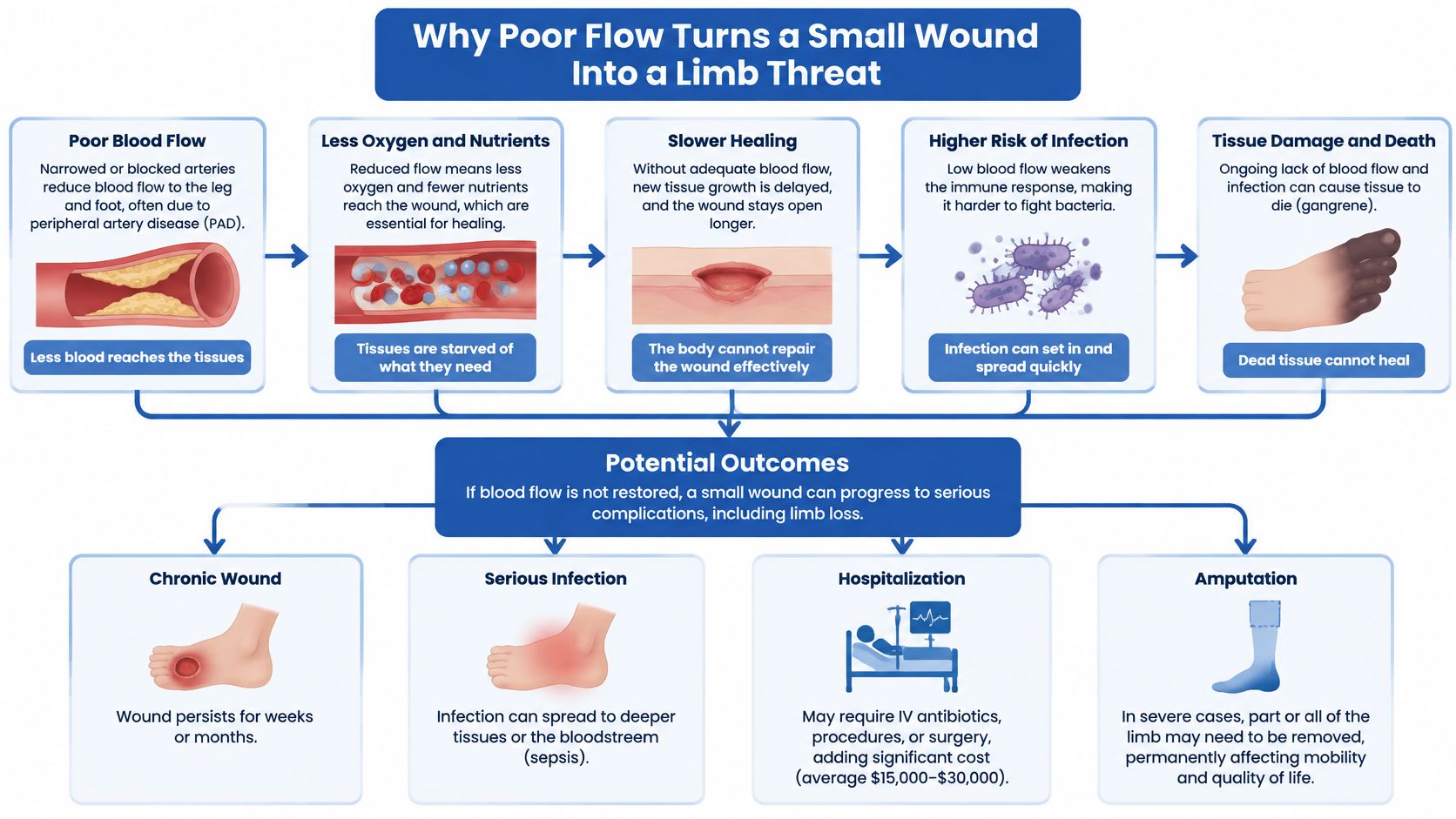
A dressing cannot replace blood flow. Call for care the same day if you see spreading redness, new drainage, odor, darkening tissue, or fever. Those are infection signs, not part of healing.
The modeled amputation gap is the practical lesson. Nerve damage can hide the wound while vessel damage stalls its repair, so the two have to be measured together, and a normal ankle-brachial index on its own does not close the question. Early detection, pressure relief, vascular assessment, and follow-up change outcomes. Waiting for the wound to look dramatic does not.
Blood Flow Tests and the Numbers That Matter
Find your number first, then read what it can and cannot tell you. Diabetes often calcifies ankle arteries, so the most familiar test is also the easiest to misread.
| Test | What it measures | Concern threshold | Limitation in diabetes |
|---|---|---|---|
| Ankle-brachial index (ABI) | Ankle pressure divided by arm pressure | Below 0.4 prompts urgent revascularization review; a noncompressible high reading is unreliable | Falsely reassuring when arteries are calcified |
| Toe pressure (toe-brachial index) | Blood pressure in the toes | Below 30 mmHg signals severe ischemia | Needs interpretation alongside other tests |
| Ankle pressure | Pressure at the ankle | Below 50 mmHg prompts urgent revascularization review | Limited when vessels are calcified |
| Transcutaneous oxygen pressure (TcPO2) | Oxygen reaching the skin | Below 25 mmHg signals severe ischemia; 25 mmHg or more favors healing | Needs specialist equipment |
| Skin perfusion pressure (SPP) | Pressure of blood flow in skin microvessels | 40 mmHg or more favors healing | Needs specialist equipment |
| Doppler waveform | Shape of the arterial flow signal | A flattened or dampened waveform suggests obstruction | Accuracy tracks examiner skill |
| Duplex ultrasound | Flow direction, reflux, and narrowing | Shows where reflux or blockage sits | Needs a trained operator and extra time |
Reading the rows in test order
The ankle-brachial index divides ankle pressure by arm pressure. It is the usual starting point, not the last word. A very low result pushes the plan toward urgent revascularization review. A stiff, calcified artery can push the number up and make it look safer than the foot is.
Toe pressure is the usual next step when the ankle reading is hard to trust. The 2019 IWGDF peripheral artery disease guideline notes that a toe pressure of at least 30 mmHg, a TcPO2 of at least 25 mmHg, or a skin perfusion pressure of at least 40 mmHg increases the pre-test probability of healing by at least 25%. A toe pressure below 30 mmHg or a TcPO2 below 25 mmHg signals severe ischemia and should prompt urgent vascular imaging and consideration of revascularization.
Ankle pressure below the urgent-review line belongs in the same conversation, especially when the ankle vessels are calcified and the index is muddy. Skin oxygen and skin perfusion pressure need specialist equipment, so they are not driveway tests. They answer a different question from the ankle index. Is oxygen really reaching skin?
A Doppler waveform is the shape of the pulse signal. A flattened or dampened wave suggests obstruction even before you memorize a ratio. Duplex ultrasound maps direction, reflux, and narrowing. A vein consultation and duplex ultrasound help identify where abnormal reflux is occurring and which treatment is most appropriate when swelling or a gaiter-area wound suggests a vein problem as well as an artery problem.
Every diabetic lower-leg ulcer gets a documented vascular assessment in the same plan as infection checks, pressure relief, debridement, and glucose management.
Ask for toe pressure or TcPO2 whenever a leg ulcer is present, including when the ankle index looks acceptable. One comfortable number should not close the chart.
Telling Arterial, Venous and Nerve Wounds Apart
A foot ulcer that barely hurts, a toe wound that aches at night, and a weeping sore above the ankle can all land in the same exam room. They look like variations of one problem. They are three problems, and each one stalls for a different reason: nerve damage hides the injury, artery disease starves the tissue of oxygen, and vein disease floods the tissue around it.
Patients ask a fair question: "What should happen when my ABI is normal, but my diabetic wound is not healing?" The answer is a second measurement, taken a different way. Diabetes-related medial arterial calcification can stiffen ankle arteries and falsely inflate the reading. The 2025 ACC/AHA scientific statement notes that ABI sensitivity is limited when vascular calcification is present and recommends a toe-brachial index when the ABI is noncompressible (above 1.40). Exercise ABI testing can also uncover disease when walking symptoms persist despite a normal or borderline resting ABI.
Sorting the wound starts with where it sits, how it looks, and what the foot feels like. An evaluation can help determine the severity of chronic venous disease, which is a different job from proving an artery is open.
| Wound type | Typical location and look | Key clues | Main treatment direction |
|---|---|---|---|
| Neuropathic diabetic ulcer | Pressure points on the sole or toes, often painless, callus around the edge | Near-normal pulses, reduced sensation | Offloading, debridement, infection control, glucose management |
| Arterial (ischemic) ulcer | Toes, heels, or outer ankle, pale or dusky dry base | Cold foot, weak pulses, pain at rest, low toe pressure | Urgent vascular assessment and revascularization review |
| Venous leg ulcer | Gaiter area above the ankle, shallow, weeping | Swelling, varicose veins, skin discoloration, heaviness | Compression after a vascular check, plus treatment of venous reflux |
| Mixed wound | Often on the forefoot or lower leg | Diabetes with low pressures and loss of sensation | A combined vascular plan and an offloading plan |
Why the label changes the plan
A neuropathic ulcer on the ball of the foot, ringed by callus and barely painful, needs pressure off the spot. A pale ulcer on a toe with rest pain needs blood flow restored. A shallow, weeping ulcer above the ankle, with heavy legs and skin staining, is the picture discussed in this comparison of venous leg ulcers and arterial leg ulcers. Mixed wounds carry both a low-flow problem and a pressure problem, so one treatment family will not cover them.
Applying compression to a leg with significant arterial disease can worsen ischemia, which is why the vascular check comes before the wrap. Once the arteries are confirmed open, compression becomes one of the most useful tools against a venous ulcer.
Non-healing wounds, abnormal pulses, or pain when you walk warrant a clinical evaluation. Do not start tight wraps on your own, and do not assume glucose is the only lever left.
What to Do When a Diabetic Wound Stops Healing
A diabetic wound that stops shrinking is reporting a missing input, and the visit's job is to find which one. Six checks, in order, separate a supply problem from a pressure problem from an infection problem.
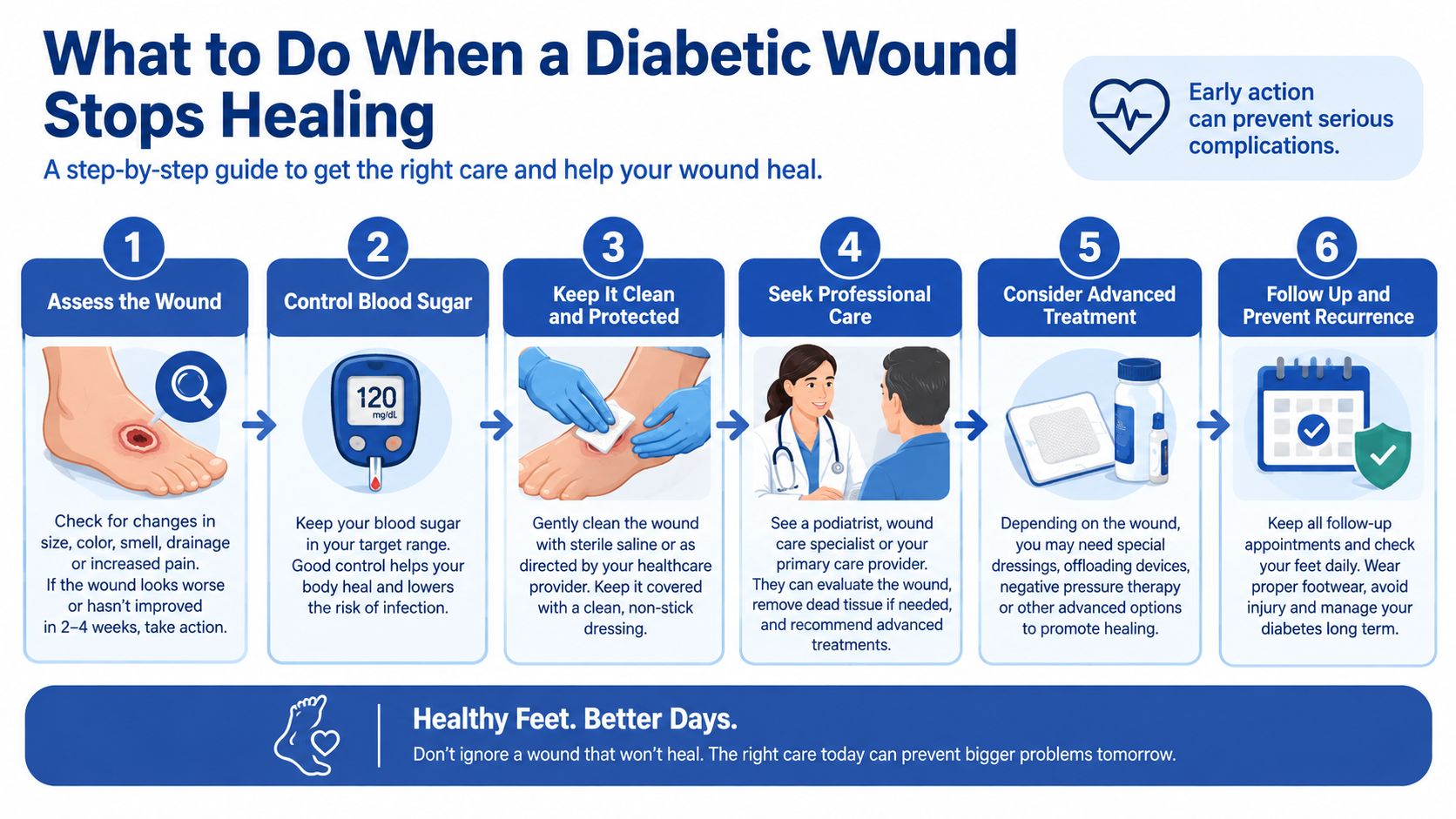
- Measure and photograph. Record length and width the same way each visit, and keep a photo. Memory is a poor ruler, and a stall is invisible without a trend.
- Check for infection. Look for redness, drainage, odor, and fever, and send cultures when the wound or the foot looks infected. Infection plus poor flow carries a markedly higher risk of poor healing and amputation, so this is not a "watch it for a month" item.
- Confirm offloading. A total contact cast, a removable walker, or padded footwear must reliably keep pressure off the ulcer. A device in the closet does not count.
- Review the glucose trend. Use home logs and clinic results. Sustained high glucose slows the same repair steps the wound is waiting on.
- Repeat the vascular assessment. Recheck pulses, and pair the ankle index with toe pressure or TcPO2, because calcified diabetic arteries can push the ankle number upward and make it read better than the foot's actual flow. A reassuring ankle index does not close the question if the toe pressure disagrees, and a result in the severe-ischemia range set out in the blood flow testing section moves the case to an urgent vascular referral.
- Use the four-week trigger. The 2023 IWGDF PAD guideline recommends reassessing vascular status and consulting a vascular specialist when a diabetic foot ulcer does not achieve a meaningful response, illustrated as less than a 50% reduction in wound area within 4 weeks, despite infection control, glucose management, wound care, and pressure offloading.
Why local care stalls
Persistent ischemia impairs inflammatory-cell function, collagen synthesis, angiogenesis, and resistance to infection. Each of those repair jobs runs on oxygen that only blood can deliver. Another ointment cannot supply those. Escalate on a stalled area reduction. Do not wait for black tissue or exposed bone to prove the point.
What Treatment Looks Like When Flow Is Low
Once reduced flow is confirmed, the next step is mapping it rather than guessing again. Duplex ultrasound usually comes first because it shows where flow speeds up, slows, or stops without any incision. When a procedure looks likely, the team typically adds a contrast angiogram, most often CT or MR based, to show how long the blockage runs, how wide the channel still is, and whether the vessels below it are worth reaching.
Restoring arterial flow
Revascularization is considered urgently once perfusion falls into the severe range used to flag ischemia. There is no single procedure with a single name. For a short, focal narrowing, an endovascular approach threads a catheter into the artery and inflates a small balloon to widen the segment, sometimes leaving a metal scaffold, called a stent, to hold it open. For a longer or more scattered blockage, surgical bypass attaches a graft above and below the diseased stretch so blood detours around it. The choice follows where the disease sits, how far it extends, your other surgical risks, and whether a usable vessel remains on each side.
Better flow restores supply; it does not by itself close the wound. Offloading, debridement, infection control, and glucose management continue until healing finishes, because a shoe that keeps pressing and dead tissue that stays put will stall repair even in a reopened artery.
Veins, mixed wounds, and the recheck
Compression is used only after a vascular check shows the leg can tolerate it. For a venous ulcer, compression pairs with treatment of the reflux itself: a small catheter or access point can close the problem vein from within so blood reroutes through healthier veins. A minimally invasive option enters the picture when compression, elevation, and activity changes have not given enough relief, and the method is matched to vein size and location, the ultrasound findings, symptoms, and overall vascular health. Afterward, patients are often asked to wear compression for a set period, avoid strenuous exercise briefly, and follow bathing or wound-care directions that vary by procedure.
For a mixed wound, the arterial plan and the offloading plan run together, and neither substitutes for the other. Judge progress by whether the ulcer is clearly smaller at the scheduled recheck, while keeping two measurements in view at once: the nerve exam that shows whether you can feel the foot, and the flow numbers that show whether it is supplied. A stall is a reason to retest both, not to wait and see.
Prevention and Coordinated Care That Protects Limbs
Prevention works like a smoke alarm plus a repair crew. Nerve damage can muffle the alarm, and vessel damage can keep the crew from arriving. That is why skin checks, pulses, and nerve sensation belong in the same routine. A normal ankle-brachial index can be reassuring and still incomplete: it may leave stiff arteries, toe-level flow, or early skin breakdown needing a closer look.
Daily habits that catch the hidden sore
- Inspect both feet every day. Use a mirror, or ask a family member, for the sole and the spaces between toes.
- Check the inside of each shoe. A rough seam or a pebble is enough to open skin you cannot feel.
- Skip barefoot walking. Even at home, a small object on the floor can start an ulcer.
- Moisturize the skin, not the toe webs. Dry cracks split. Damp webs grow infection.
- Stop smoking. Tobacco narrows arteries that diabetes is already injuring.
Glucose, blood pressure, and cholesterol targets are set with your clinician rather than borrowed from a neighbor. An annual foot and pulse exam belongs on that same calendar, and sooner when callus, redness, swelling, or a new sore appears. Short movement breaks help the legs you are trying to protect. These exercise ideas for adults over 50 are a practical place to start when a long workout is not realistic.
Screening before classic leg pain
Do not let the absence of calf pain set the schedule. In diabetes, neuropathy can blunt the usual walking pain, so age, foot complications, microvascular disease, or kidney and eye damage are reasons to measure circulation even when walking feels normal. The practical question is simple: can this foot feel injury, and can blood reach the spot fast enough to repair it?
Access shapes the outcome as much as the biology. Where vascular testing and referral are scarce, peripheral artery disease can stay invisible until a wound stops healing. The American Heart Association, writing in JACC, reports that Native American, Black, Hispanic, rural, and low-income populations are more likely to be uninsured, with lack of insurance independently associated with amputation risk. Diabetes combined with PAD produces more complications than either condition alone.
A wound can worsen because vascular testing or specialist referral happened too late, even when diabetes care is otherwise active. Insurance gaps, transportation, language, and education that does not fit the person all slow that referral. Primary care, wound care, and vascular evaluation work best as one sequence. Vein & Wound Institute brings advanced vein care and specialized wound management together under one roof, so reflux and an open wound are not booked as unrelated problems. Day-to-day protection is spelled out in this guide on how to prevent wounds and ulcers if you have diabetes.
Book a Circulation Check If Warning Signs Appear
Book an evaluation if you have diabetes and any of these show up. Do not wait for the wound to look severe.
- A foot or leg wound that has barely shrunk after several weeks of real offloading, wound care, and glucose attention.
- A cold, pale, or dusky foot, or pain at rest, or calf pain that stops you from walking.
- Visible or bulging varicose veins, leg aching, heaviness, or fatigue.
- Swelling related to venous insufficiency, or skin changes and complications near the ankle from chronic venous disease.
A minimally invasive procedure may be considered when venous disease causes persistent symptoms or when compression, elevation, and activity changes do not provide enough relief. Diabetic and non-healing wound care is focused on healing, infection prevention, circulation, and long-term foot and leg health, so the visit is about the limb, not a single dressing change.

Bring a glucose log, your current medication list, wound measurements and photos, and any earlier ankle index or imaging. Ask whether toe pressure or skin oxygen has been measured, whether duplex ultrasound is needed, and who will coordinate with your primary care clinician. We explain the findings in plain language and align the plan with that clinician. A normal ankle index, on its own, is not a reason to wait.
Vein and Wound Institute evaluates circulation and treats diabetic and non-healing wounds in one coordinated plan, including the vein problems that keep leg ulcers open. If a sore is stalling, or your legs ache, swell, or show skin changes, schedule a visit with Vein and Wound Institute and bring your wound photos and any prior flow tests.