
Could a brown patch on your lower legs be more than a surface stain? Marks around both ankles often reflect venous pressure, but appearance alone cannot confirm the cause. The color can remain after pressure has changed, like a rust stain left after a leak is repaired.
Swelling, itch, or fragile skin alongside the discoloration can offer useful clues. A patch that does not hurt may still deserve attention; judging urgency depends on where it appears, what other symptoms accompany it, and whether it is new, spreading, painful, or open.
For example, rust-colored marks above both ankles after a week on your feet may seem like an ordinary sign of aging. Their location and any accompanying changes matter more than color alone. A brown mark is a reason to check, not a diagnosis.
Table of Contents
- What Brown Splotches on Legs Usually Mean
- Causes at a Glance and How to Tell Them Apart
- How Venous Pressure Leaves Brown Marks
- Post-Inflammatory and Other Skin Causes
- Venous Warning Signs and When to Seek Care
- Will the Brown Fade and Does Skin Tone Change It
- Get a Venous Evaluation Before Treating the Spots
What Brown Splotches on Legs Usually Mean
After a long shift, you notice a rust-colored patch above your ankle. It may look like a surface stain, but its color can record what happened beneath the skin. Brown splotches on legs can form when sustained pressure in the leg veins allows small amounts of blood to seep near the skin. As red blood cells break down, iron-containing pigment remains, leaving a brown or rust-colored mark. A stain can outlast the leak that made it.
The mark's appearance offers clues, not a diagnosis. A flat tan patch may follow a bite, scrape, or rash. A scaly ring can suggest a fungal infection, while an itchy patch may appear where a product touched the skin. Pain, an ulcer, or a cold foot calls for prompt attention because these signs can occur with other conditions, including arterial disease.
To judge the likely cause and urgency, ask:
- Where are the marks? Ankles and lower calves on both legs suggest a different pattern from spots on the shins, one foot, or an old scar.
- What else do you notice? Swelling, heaviness, itch, and visible varicose veins add useful context.
- What has changed? New or spreading marks, pain, or open skin deserve faster assessment than a stable patch.
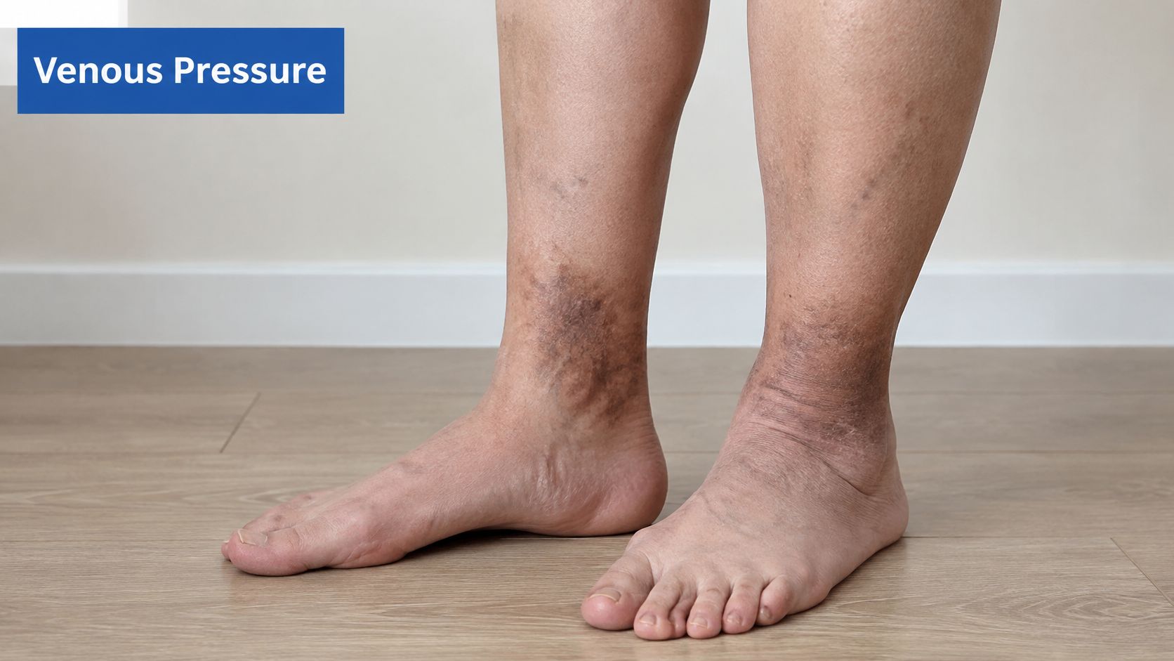
On darker skin, color changes may be less obvious or appear in different shades, so location and changes in texture or symptoms can be just as helpful as color. Use these observations to guide what to check next; testing may be needed to identify the cause.
Causes at a Glance and How to Tell Them Apart
Location comes first. Companion signs come second. One good match is a lead, not a diagnosis, and venous staining can sit on top of an older injury mark.
| Cause | Typical appearance | Where it appears | Companion signs |
|---|---|---|---|
| Venous stasis dermatitis | Rust-brown, pinpoint to confluent patches | Ankles and lower calf, both legs | Swelling, varicose veins, itch, skin thinning or thickening |
| Post-inflammatory hyperpigmentation | Flat tan to dark brown patches matching an old injury | Anywhere the skin was hurt | Usually none once the trigger has healed |
| Pigmented purpuric dermatoses | Red-brown pinpoint spots that do not fade when pressed | Shins, often both legs | Sometimes itch; usually no swelling |
| Diabetic dermopathy | Light brown, round or oval, scar-like patches | Shins | Usually painless; linked to diabetes |
| Contact dermatitis | Brown patch with redness or scaling | Where a product touched the skin | Itch; recent new product or plant contact |
| Fungal infection (tinea) | Scaly, ring-shaped, patchy brown | One leg or the feet | Itch, scale at the edges |
| Vasculitis or arterial disease | Brown or purple areas, sometimes with ulcer | Lower legs or feet | Pain, cold feet, ulcer; needs prompt review |
How to read a match
Start with the map on your own legs. Rust that begins at both ankles and creeps up the lower calf fits venous staining better than a round shin spot that looks like a faded scar. Press a pinpoint red-brown spot. If it stays colored, think of pigmented purpuric change rather than a bruise that should blanch.
Then add what you feel. Ankle swelling by evening, twisted veins, and itch pull toward veins. A new lotion and a matching itchy patch pull toward contact. Scale at the edge of a ring on one foot pulls toward fungus. Pain, a cold foot, or an open sore pulls you out of the "watch it" column.
Two causes can be true at once. An old scrape can darken on a leg that also has venous pressure, so clearing the injury story doesn't cancel the circulation story. If the row you match includes pain, ulcer, or a cold foot, don't wait on a home comparison.
How Venous Pressure Leaves Brown Marks
How does pressure in a leg vein turn into a lasting brown mark? The change happens in stages: blood pools, tiny vessels leak, and iron-containing pigment remains in the skin.
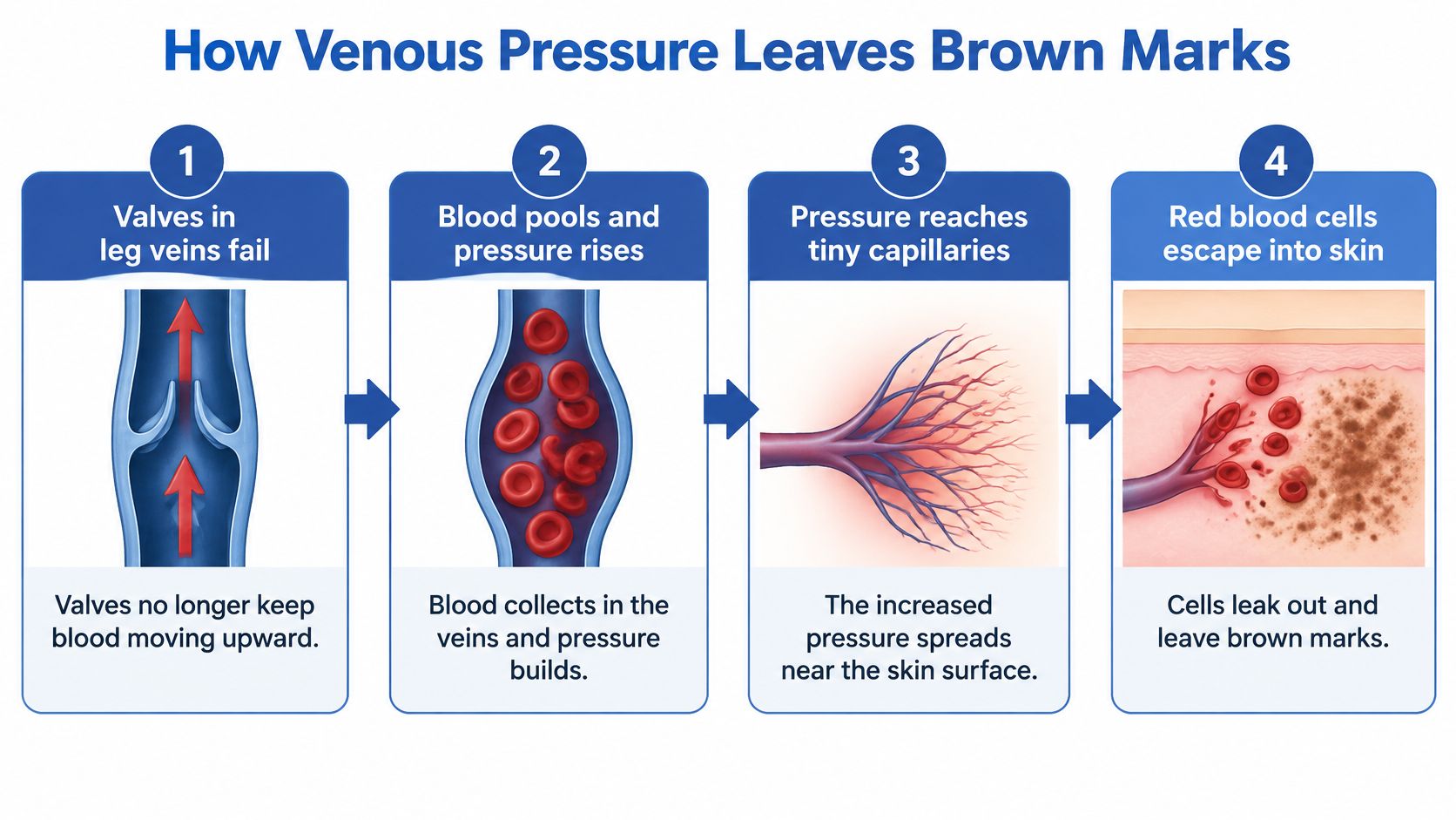
The leak, then the stain
Leg veins use one-way valves to move blood upward. When valves fail, blood can pool and pressure rises. The added pressure reaches tiny capillaries near the skin, allowing red blood cells to escape into surrounding tissue. Immune cells break down the cells' hemoglobin, and iron left behind collects as hemosiderin. Inflammation, and sometimes extra melanin, can deepen the rust or brown color. This chain is associated with chronic venous insufficiency, a circulation problem rather than pigment sitting on otherwise healthy skin.
Gravity concentrates pressure lower in the leg, so the gaiter area, around the ankle and lower calf, often shows changes first. Specks may spread into patches and merge into a broader brown area that can reach the feet. The pattern can offer clues, but it cannot show how much underlying venous disease is present.
Why the color can outlast the pressure
Skin affected by chronic venous pressure may swell, itch, dry, redden, thin, or thicken. Some people develop ulcers. A review of venous stasis dermatitis estimates that 2–6 million Americans have advanced chronic venous insufficiency with swelling or skin changes, while approximately 500,000 have venous leg ulcers. It also notes that pigmentation may persist after circulation improves because hemosiderin remains in the skin.
Treating the pressure can limit further leakage, but it does not remove iron already deposited in the skin. The mark records past pressure; its color alone cannot tell whether the pressure is still active. Swelling, skin changes, and other symptoms help provide context, while an evaluation can assess the underlying circulation.
Post-Inflammatory and Other Skin Causes
The common mistake is to name every brown patch stasis dermatitis, or to call every red ankle an infection. Both guesses cause harm. A venous mark treated as cellulitis can bring antibiotics you don't need. An infection or an arterial problem treated as a harmless stain can delay care that should have started the same day.
Post-inflammatory hyperpigmentation is the frequent non-venous pattern. A bug bite, scrape, burn, rash, or inflamed pimple heals, and a flat tan to dark brown patch keeps the shape of that injury. Many of these marks fade over months. On darker skin they often linger longer. People comparing acne-related marks sometimes look at ingredients for stubborn acne marks to see which brighteners are discussed for that kind of leftover pigment. Leg skin with swelling is a different problem, so don't borrow a face routine and skip the leg exam.
Marks that mimic a vein stain
Other patterns deserve their own names.
- Pigmented purpuric dermatoses: pinpoint red-brown spots, often on both shins, that don't fade when pressed, usually without swelling.
- Diabetic dermopathy: light brown, round or oval, scar-like shin patches that are usually painless.
- Contact dermatitis: a brown, red, or scaly patch exactly where a product or plant touched.
- Fungal infection: a ring with scale at the edge, often on one leg or the feet, plus itch.
- Medications and other inflammation: some drugs and inflammatory disorders darken skin without a vein story.
- Vasculitis or arterial disease: brown or purple areas plus pain, ulcer, or a cold foot.
The Merck Manual notes that a visual diagnosis is often appropriate, while duplex ultrasound helps when the picture is unclear or reflux or obstruction is suspected, and an ankle-brachial index matters when arterial disease is possible. It also draws the infection line clearly. Cellulitis is more often one leg, with acute tenderness and sometimes fever or other systemic symptoms. Stasis discoloration is commonly bilateral and chronic.
Ask two questions before you pick a label. Does the patch match an old injury? Does it itch and carry scale? Yes to the first leans post-inflammatory. Yes to the second, on one limb and without swelling, leans fungal or contact. Pain, heat, or fever means you stop guessing.
Venous Warning Signs and When to Seek Care
Spend one to two weeks noting what your legs do, not only how they look. Tick what repeats.
- Heaviness or aching that builds through the day: worse after standing or sitting, easier after you lie down.
- Ankle swelling that eases overnight: socks leave deeper marks by evening.
- Bulging or twisted veins: visible ropes, not only spider-fine threads.
- Itch around the ankles: often with dry or shiny skin.
- Skin that looks tight or thickened: a change you can feel, not only see.
- Brown marks on both legs, plus history: varicose veins in the family, past pregnancies, long standing or sitting, or an earlier clot or leg injury.
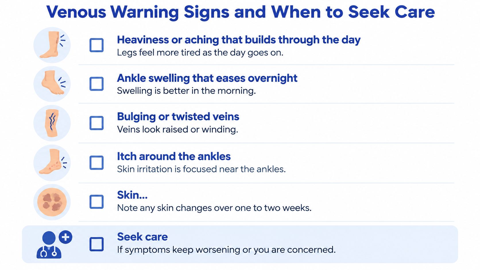
Several of these together raise the chance of venous disease. None of them proves it alone. Published estimates place chronic venous insufficiency at approximately 10%–35% of adults in the United States, and skin-pigmentation changes in the general population have ranged from 3% to 13%, according to a clinical review of chronic venous disease. Similar pigment can still come from skin disease, inflammation, medication, or a systemic condition, so a common pattern isn't a closed case.
How fast to act
| Warning sign | What it may mean | Suggested timing |
|---|---|---|
| Open sore, weeping or crusted ulcer on the ankle | Venous or arterial ulcer risk | Specialist review within days |
| Warm, red, tender, spreading area, possibly with fever | Possible cellulitis or infection | Same-day medical care |
| Pain at rest, cold or pale foot, numbness | Possible arterial insufficiency | Urgent or emergency care |
| One leg only, sudden swelling with calf pain | Possible deep vein clot | Emergency care |
| Brown marks on both legs with ankle swelling, heaviness or varicose veins | Probable venous disease | Routine vein evaluation |
| Single itchy scaly patch with no swelling | Possible fungal or contact cause | Primary care or dermatology |
Symptoms that may lead to treatment include visible or bulging varicose veins, leg aching, heaviness or fatigue, swelling related to venous insufficiency, and skin changes tied to chronic venous disease. Warning signs that varicose veins have become dangerous are worth reading beside that list, because color alone isn't the danger line.
The American Academy of Family Physicians describes rusty-red splotches in the gaiter area as hemosiderin staining, more meaningful when paired with dependent edema, varicose veins, itch, skin thickening, or a medial-ankle ulcer. For chronic bilateral edema or suspected reflux, duplex ultrasonography with reflux testing can confirm insufficiency. An ankle-brachial index is useful when arterial disease is possible before strong compression is prescribed. A vein consultation and duplex ultrasound identify where abnormal reflux is occurring and which treatment fits. Clinics that document vascular visits, including tools such as PatientNotes for vascular surgeons, show why a written symptom timeline helps the next clinician.
Don't start firm compression on your own if a foot is cold, pain comes on at rest, or arterial disease is possible.
Seek prompt assessment if discoloration comes with an open sore, drainage, rapidly increasing swelling, severe pain, warmth, or fever.
Will the Brown Fade and Does Skin Tone Change It
Will brown marks fade when the underlying vein problem improves? Sometimes, but the skin may keep a record of past pressure. Hemosiderin, inflammation, and extra melanin can remain even after venous pressure is controlled. A review of venous skin changes across skin tones highlights why appearance and recovery can differ. Better flow may help prevent worsening and ulcers, while pigment changes fade slowly or remain.
Symptom relief and pigment reduction are separate goals. Many people notice vein symptoms ease during the first several weeks after treatment, but that does not mean the brown color will clear on the same schedule. The marks may lighten partly, or barely at all.
Hue is a weak guide
Photographs taken on lighter skin teach the wrong template. On darker skin, the same process may appear brown, purple, gray, or ashen. The familiar red-to-brown speckling can be subtle or absent, so a lack of textbook color does not rule out venous skin changes.
| Skin tone | Typical color of marks | What to watch instead |
|---|---|---|
| Lighter skin | Red-brown to rust-brown | Classic visible signs such as pinpoint rust staining |
| Darker skin | Brown, purple, gray or ashen | Subtler visual signs; itch, heaviness, tightness and recurring wounds matter more |
Itch, heaviness, swelling, tightness, and recurring wounds offer clues that color alone cannot. Ask for an exam that accounts for your skin tone, and mention it if the photographs in the room do not reflect how your skin looks. The pattern of symptoms and a full assessment matter more than matching a picture.
Topical treatments may help some superficial dark spots, but they do not address venous pressure or remove iron pigment. Discuss any topical plan and daily sun protection with a clinician, since fragile skin may react to irritating or unproven bleaching mixtures. Information about brightening ingredients for dark spots can provide cosmetic context, not treatment for the underlying vein problem.
Get a Venous Evaluation Before Treating the Spots
A useful visit starts with timing. When do the legs ache, when does swelling peak, and what makes both ease? The exam then looks for swelling, varicose veins, and skin change, including thickness and any break in the skin. Duplex ultrasound maps abnormal reflux. An ankle-brachial index comes in when arterial disease is possible. If the skin diagnosis stays unclear, a biopsy may be the next step rather than another cream.
Match the clinician to the picture.
- Vein specialist: suspected reflux, bilateral ankle staining, heaviness, or varicose veins.
- Dermatology: rash, fungus, contact, or pigment that doesn't fit a vein map.
- Wound care: any open sore.
- Primary care: the place that sorts referrals when more than one of these is true.
An evaluation can help determine the severity of chronic venous disease. Bring four questions: What is causing the marks? Which tests are needed? What compression level is appropriate, and for how long? What should I expect for the pigment versus the circulation?
The treatment plan is based on the source of the problem, not on the veins visible at the skin's surface. When an unhealthy vein is closed or removed, blood redirects through healthier veins, which can lower pressure and ease symptoms. A minimally invasive procedure may be considered when symptoms persist or when compression, elevation, and activity changes don't give enough relief. The choice depends on vein size and location, ultrasound findings, symptoms, and overall vascular health.
Vein and Wound Institute offers that duplex-based evaluation together with wound care when skin has already broken. If you are unsure who should see you first, this guide on how to choose a vein care specialist lays out the decision. If the warning signs above match your week, or the symptoms have stayed despite elevation and movement, book the evaluation rather than waiting on the color to decide for you.
Vein and Wound Institute evaluates chronic venous insufficiency and the skin changes that come with it, including brown lower-leg discoloration, using clinical examination and duplex ultrasound across Los Angeles, Santa Barbara, and Ventura Counties. If your ankles show persistent staining with swelling, heaviness, or a slow-healing spot, visit Vein and Wound Institute to schedule an evaluation and sort circulation from pigment before you treat the surface.