
A sore above the ankle that keeps reopening can make every dressing change feel like a setback. You may be following the wound-care instructions and wearing compression, yet the skin still isn't closing. That doesn't necessarily mean you're doing something wrong. A venous ulcer can stall when the pressure causing it hasn't been fully identified, or when the compression plan doesn't fit the leg's arterial blood supply and vein findings.
The first step is to understand what the sore is showing you. Then clinicians can assess circulation, choose safe wound care, and decide whether treating a problem vein could help.
Table of Contents
- What a Venous Leg Ulcer Is and How Common It Is
- How Venous Reflux Turns a Leg Into an Open Wound
- Diagnosing the Cause With Duplex Ultrasound
- Staging Your Ulcer by Size, Duration and Healing Odds
- Wound Care and Compression Done Safely
- Vein Treatments That Close the Source of Reflux
- Compression After Healing and Preventing Recurrence
- Why an Ulcer Stalls and When to Book Review
What a Venous Leg Ulcer Is and How Common It Is
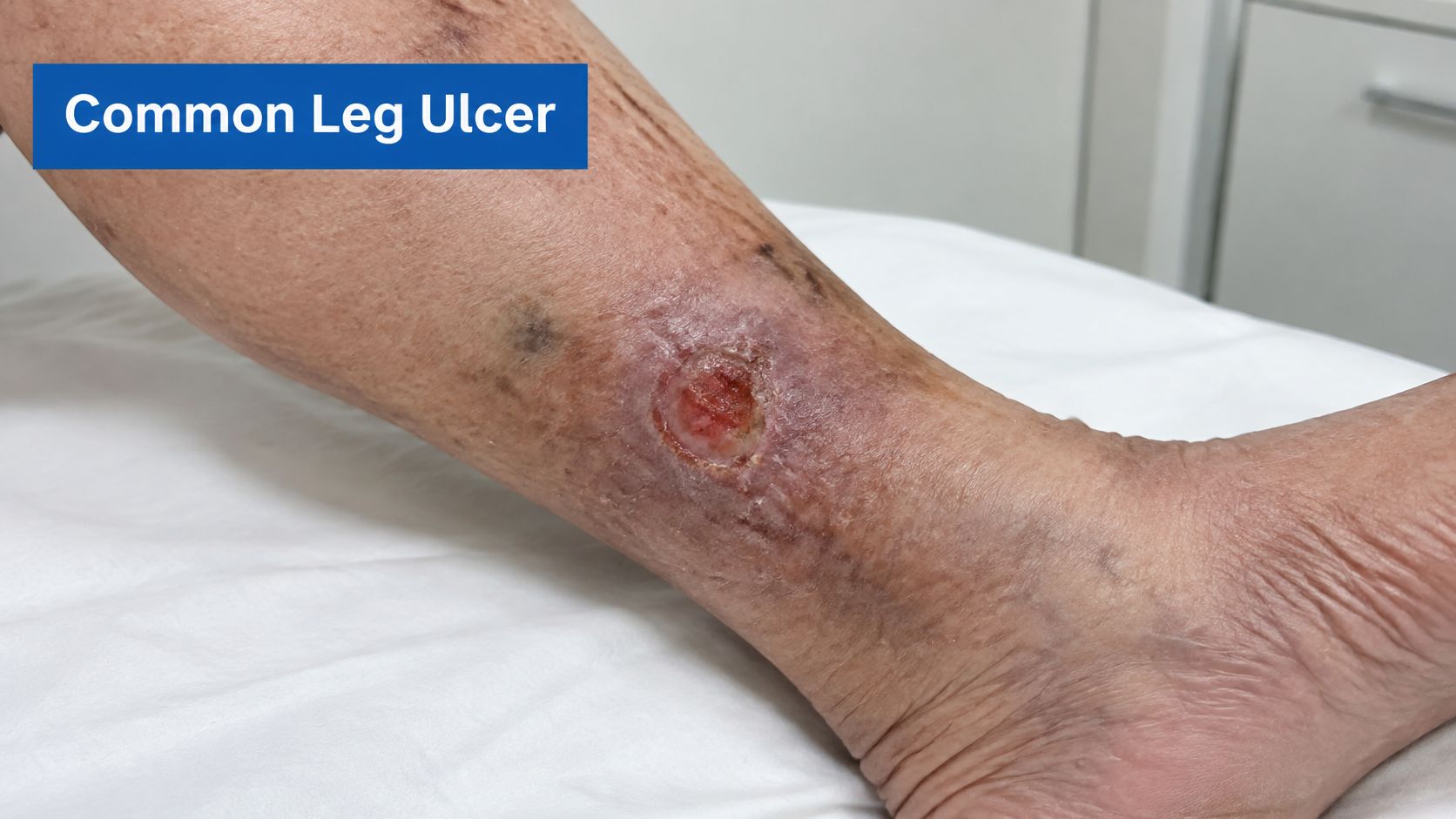
An open sore above the ankle that does not heal may be a venous leg ulcer (VLU). It develops when blood does not return efficiently toward the heart, raising pressure in the leg veins. The circulation problem behind it is called chronic venous insufficiency. The ulcer is a visible sign of that problem, so wound care needs to account for the circulation affecting the skin.
How common are venous ulcers?
Venous ulcers account for approximately 60% to 80% of leg ulcerations associated with venous disease, according to a review of the condition. A 2023 systematic review and meta-analysis that pooled 13 studies estimated global prevalence at 0.32%, with a 95% confidence interval of 0.129% to 0.595%, and pooled incidence at 0.17%. (The 2023 review of venous leg ulcer prevalence)
Estimates differ because studies may define ulcers differently, include populations with different age structures, or count active and previously healed ulcers in different ways. For an individual patient, the location and appearance of a sore are clues, but they do not by themselves establish its cause.
In the United States, clinical reviews commonly estimate that chronic venous insufficiency affects 10% to 35% of adults, while venous ulcers affect roughly 1% to 3% of the population. About 4% of adults aged 65 and older are reported to develop them. A 2024 systematic review and meta-analysis estimated that venous leg ulcers affect up to 2% of the population. (The 2024 venous leg ulcer review)
A venous ulcer may remain open even when dressings are changed and compression is used. Skin care alone does not correct the venous pressure driving tissue breakdown. Compression strength must suit the leg's arterial blood supply, and decisions about vein treatment depend on the source of reflux. A clinician assesses those factors to guide care, rather than relying on one standard approach for every ulcer.
For a plain-language comparison of arterial and venous ulcers, their causes, symptoms and treatment, see this guide.
How Venous Reflux Turns a Leg Into an Open Wound
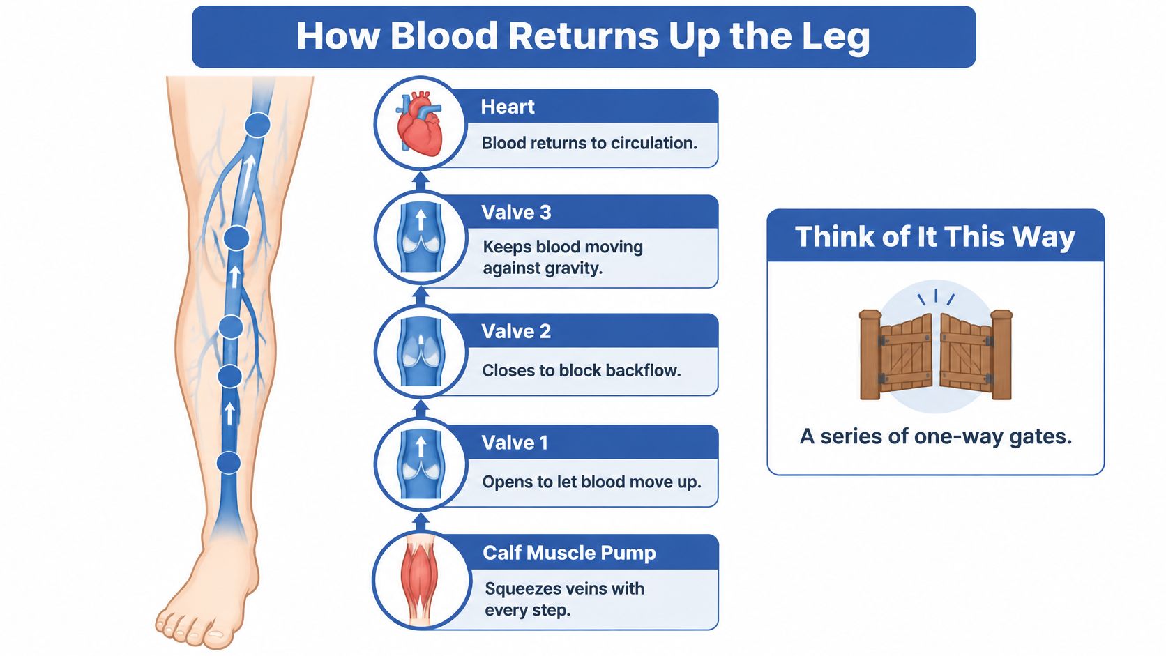
Healthy leg veins return blood upward against gravity. One-way valves help keep blood moving in the right direction, and the calf muscles squeeze the veins when you walk. Together, the valves and muscles help push blood back toward the heart.
Think of the valves as a series of one-way gates on a stairway. Each gate opens to let blood move upward and closes to limit backflow. If a gate fails and swings the wrong way, blood can slip back down and crowd the veins below it.
From leaking valves to skin breakdown
Venous reflux means blood flows backward through a vein valve that isn't closing properly. When blood pools, especially during prolonged standing or sitting, pressure in the veins can stay too high. This sustained pressure is called venous hypertension.
Over time, the pressure stretches small veins and capillaries, the tiny blood vessels that connect arteries and veins. Fluid and proteins can leak into surrounding tissue, causing swelling and inflammation. The skin may become darker, thicker, or firmer before it breaks down into an ulcer.
Reflux can arise in superficial veins, which lie close to the skin, including the great saphenous vein. It can also involve perforator veins, which connect superficial veins to deeper veins. Deep vein disease or an obstruction to blood flow may contribute as well, so the visible varicose veins don't always reveal the full cause.
Why the vein assessment matters: Closing the wound treats the damaged tissue. It doesn't necessarily remove the pressure that caused the damage.
That's why a wound can improve and then reopen if the underlying venous problem remains. A broader introduction to chronic venous insufficiency, its symptoms, causes and treatment can help put the circulation problem in context.
Diagnosing the Cause With Duplex Ultrasound
A leg ulcer may remain open even with dressings and compression if the treatment does not match the circulation problem. The right compression strength depends in part on arterial blood flow, while any vein treatment depends on where reflux begins. An evaluation can help assess the severity of chronic venous disease and guide those decisions.
| Test | What It Reveals | When It Is Used |
|---|---|---|
| Clinical examination and history | Wound edges, skin changes, varicose veins, swelling, previous ulcers and pain pattern | At the first visit |
| Ankle-brachial pressure index (ABPI) | Compares blood pressure at the ankle and arm to assess arterial blood flow | Before strong compression is considered |
| Duplex ultrasound | Direction of blood flow, reflux in superficial, deep and perforator veins, and possible deep vein blockage | To locate abnormal reflux and assess veins before treatment |
| Wound swab or biopsy | Possible infection or unusual tissue causes | If a wound looks infected, unusual or slow to heal |
| Blood tests | Possible diabetes, anaemia, low albumin or nutritional deficits | If slow healing or another health concern is suspected |
The order of assessment
The clinician starts by asking about your health history and examining the leg. They may check the wound's size and edges, swelling, visible veins and skin changes. Previous ulcers and the pattern of pain can also help distinguish venous problems from other causes.
Next, an ankle-brachial pressure index (ABPI) may be measured. This compares systolic blood pressure, the pressure when the heart contracts, at the ankle and arm. The result helps show whether enough arterial blood is reaching the foot. Strong compression can be unsafe if significant arterial disease restricts that flow, so compression strength should reflect the findings.
A duplex ultrasound combines vein images with measurements of blood flow. For example, the scan can show blood flowing backward in the great saphenous vein, a superficial vein running along the leg, and identify where the reflux starts. It can also assess deep and perforator veins and check for signs of blockage. This map helps determine whether a vein treatment may address the source of pressure, rather than targeting only veins visible at the skin.
A swab or biopsy may be considered if the ulcer appears infected or unusual, or is not healing as expected. Blood tests may identify health issues such as diabetes, anaemia or poor nutrition that can slow healing.
The sequence matters: check arterial blood flow before choosing strong compression, then use ultrasound findings, symptoms and overall vascular health to guide any vein treatment.
Staging Your Ulcer by Size, Duration and Healing Odds
Staging helps describe both the venous disease and the wound. In the CEAP clinical classification, a system used to describe chronic venous disease, C6 means there's an active venous ulcer; a healed ulcer is classified as C5. For wound care, size and how long the ulcer has been present help estimate how difficult it may be to close.
| Scenario | Reported Outcome | Context |
|---|---|---|
| Wound under 10 cm² and under 1 year old | 29% chance of failing to close by 6 months | Prognostic model |
| Wound over 10 cm² and over 1 year old | 78% chance of failing to close by 6 months | Prognostic model |
| All venous leg ulcers | About 60% heal within 12 weeks | Widely cited evidence summary |
| All venous leg ulcers | 30% to 70% heal by 6 months; 10% to 20% remain under treatment after 1 year | Review range |
| Community-based care | About 45% healed at 6 months | Scottish Leg Ulcer Trial |
| Specialist clinic care | About 70% healed at 6 months | Historical UK guidance comparison |
| Recurrence after healing | 26% to 69% at 12 months in historical UK data; more than 60% of treated ulcers recurrent in some studies | Epidemiological estimates |
The figures in the table come from a review of venous leg ulcer outcomes and prognostic evidence. (Evidence on healing and recurrence in venous leg ulcers)
What size and duration can tell you
A smaller, newer wound generally has better healing odds than a larger wound that has been present for a long time. In a prognostic model, a wound under 10 cm² and less than one year old had a 29% probability of failing to close by six months. A wound over 10 cm² and more than one year old had a 78% probability of closure failure. Those figures describe groups, not a certain outcome for an individual.
Across reviews, about 60% of venous ulcers heal within 12 weeks. Reported healing by six months ranges from 30% to 70%, and 10% to 20% remain under treatment for more than a year. Differences in wound severity and care setting help explain why reported outcomes vary.
Why specialist review can change the plan
Historical UK guidance reported that about 45% of ulcers treated in community-based care healed by six months, compared with about 70% in specialist clinics. These comparisons don't guarantee what will happen to a particular wound, but they underline the value of reassessing an ulcer that isn't improving.
If the wound is in the higher-risk size-and-duration category, or it hasn't clearly shrunk over several weeks, ask for a vascular and wound-care review. The purpose is to check whether the original diagnosis and treatment plan still fit the wound's progress.
Wound Care and Compression Done Safely
Good dressings alone often fail when the pressure driving the ulcer stays high. Compression lowers that venous pressure, but the safe strength depends on arterial blood flow and on where the reflux starts. The plan is set for the leg in front of the clinician, not from a single standard protocol.
Before strong compression is used, arterial inflow is checked, typically with an ABPI. This is a comparison of blood pressure at the ankle and arm. Compression can worsen ischaemia, meaning inadequate blood flow to tissue, when significant peripheral arterial disease is present. A leg with suspected arterial disease needs vascular evaluation before routine high pressure treatment.
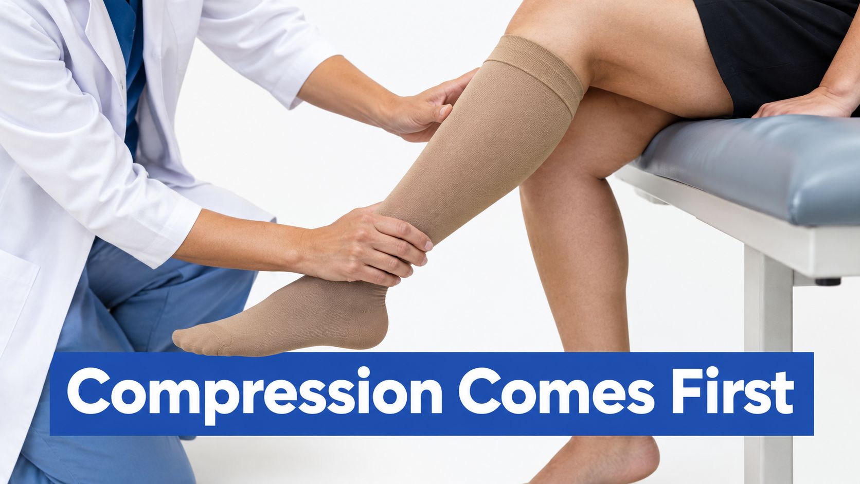
When arterial inflow is adequate, compression is the main treatment that counteracts the venous pressure behind the ulcer. Strong compression may use an elastic system delivering at least 40 mmHg at the ankle, or a properly applied inelastic system. (NICE recommendations on compression and assessment for leg ulcers) That level is a clinical treatment dose, not a direction to select or apply a bandage without supervision.
How compression supports healing
Outside pressure works like a firm layer around the calf. It limits pooling and swelling, and it helps the calf muscle pump push blood upward during walking. Better upward flow lowers venous hypertension and supports oxygen and nutrient exchange in the skin.
Several factors shape the choice. Clinicians weigh arterial status, leg shape, amount of wound fluid, mobility, pain and tolerance. Tell the clinic the same day if a bandage slips, wrinkles, or causes new discomfort. Do not tighten it yourself.
Wound care and compression are planned together. The clinician may clean the wound, remove dead tissue through debridement, choose a dressing for the level of fluid, and check for pain or infection. The dressing protects the wound and manages fluid. Compression treats the pressure underneath.
For a practical overview, see compression therapy and its role in venous care. Contact your care team for reassessment if you notice:
- Increasing pain, especially after compression is applied.
- A cool or pale foot, which may suggest reduced blood flow.
- Numbness or other new nerve symptoms.
- A slipping bandage or toes that swell.
- Persistent swelling despite the current plan.
Vein Treatments That Close the Source of Reflux
A wound may remain open even with good dressings and compression if pressure from faulty veins continues to affect the skin. For selected patients, treating superficial venous reflux while the ulcer is active can support faster healing. In the EVRA randomized trial, early endovenous treatment was associated with a median healing time of 56 days, compared with 82 days when treatment was deferred. Healing by 24 weeks was 85.6% versus 76.3%. (EVRA trial results and follow-up)
| Option | How It Works | Typical Use | Anaesthesia and Recovery |
|---|---|---|---|
| Endovenous laser ablation | Laser energy delivered through a catheter closes the vein from within | Reflux in the great or small saphenous vein | Local anaesthesia; usually walking the same day |
| Radiofrequency ablation | Heat delivered through a catheter closes the vein from within | Similar to laser; selected based on vein size and clinic protocol | Local anaesthesia; usually walking the same day |
| Foam sclerotherapy | Ultrasound-guided foam is injected to close the vein | Smaller veins and branch reflux | Outpatient treatment; compression afterwards |
| Ambulatory phlebectomy | Small incisions are used to remove bulging surface veins | Symptomatic varicose tributaries | Local anaesthesia; compression and short activity limits |
| Open surgical ligation or stripping | Incisions are used to tie off or remove the vein | Complex cases, or when endovenous treatment isn't suitable or hasn't worked | General or regional anaesthesia; longer recovery |
What vein closure means
Endovenous treatment closes an unhealthy vein from within, using a small catheter or access point. Blood then reroutes through healthier veins, which can reduce venous pressure. This differs from traditional stripping, which removes a vein through incisions.
Most minimally invasive endovenous procedures use local anaesthesia to numb the treatment area, and patients usually remain awake. Pressure or brief discomfort may occur, but significant pain is not typically expected. Recovery instructions vary by procedure, including advice about compression, activity and wound care.
The right procedure depends on the vein's size and location, ultrasound findings, symptoms and overall vascular health. Duplex mapping identifies the reflux source that surface veins can hide. It also helps clinicians assess whether a procedure is appropriate alongside wound care and compression.
Closing a vein does not remove the underlying tendency toward venous disease. In long-term EVRA follow-up, early intervention was associated with a lower recurrence incidence rate, 0.11 rather than 0.16 episodes per person-year, although the time to first recurrence was not statistically different between groups. These findings support considering reflux treatment as part of a wider care plan, with the approach adjusted to the source of the problem and the patient's vascular health.
Compression After Healing and Preventing Recurrence
Once an ulcer has closed, ask your clinician whether to continue compression. The decision depends on whether venous hypertension, meaning persistently high pressure in the leg veins, remains.
UK guidance distinguishes between cases where treatment resolves purely superficial venous hypertension and cases where raised venous pressure persists. If the pressure has resolved, long-term compression may not be needed. If it remains, compression should continue at a level that supports healing and feels manageable. (UK guidance on compression after healing and vein treatment)
Check that garments still fit and feel comfortable, and arrange review if swelling returns or skin changes appear. Set an individualized compression plan with your clinician and revisit it at follow-up. The right level depends on your circulation and the cause of the venous pressure.
Daily habits can also support leg health:
- Walk regularly to activate the calf-muscle pump, which helps move blood up the leg.
- Raise your legs while sitting to ease pooling and swelling.
- Moisturise and protect your skin to reduce dryness and the chance of injury.
- Manage weight and blood sugar where relevant as part of your overall care.
- Keep vascular and wound follow-up appointments so changes can be assessed early.
As noted earlier, an ulcer can return even after it heals. Continued follow-up and a compression plan suited to your circulation help address that risk.
Why an Ulcer Stalls and When to Book Review
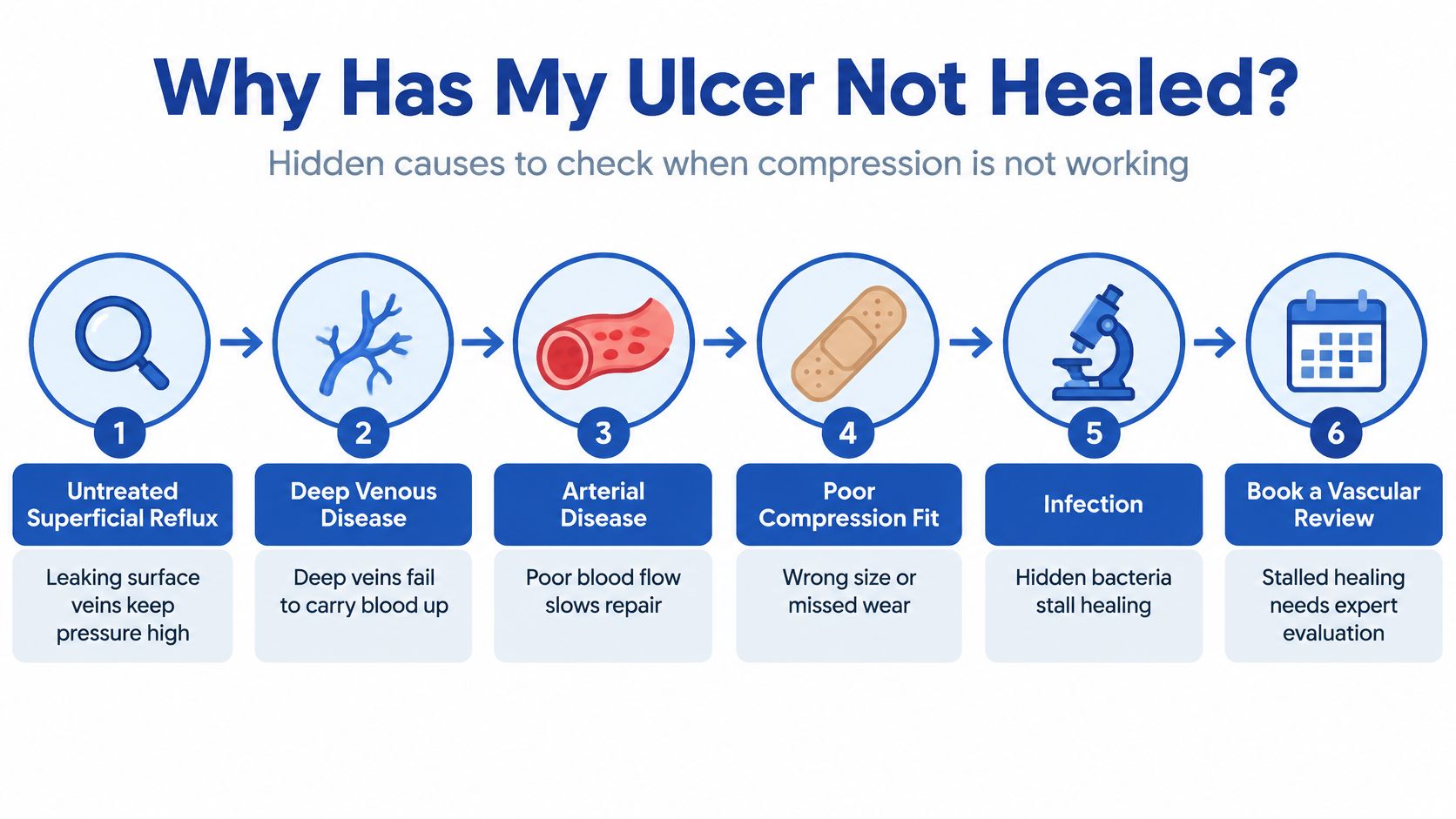
Why might an ulcer fail to heal despite compression? A common reason is that the wound care is addressing the skin while a hidden circulation problem remains. Other possible causes include:
- Untreated superficial reflux, which keeps pressure high in surface veins.
- Deep venous disease or obstruction, which affects blood return through deeper veins.
- Arterial insufficiency, meaning reduced blood flow through the arteries.
- Infection, which can interfere with healing.
- Lymphoedema, or a buildup of fluid in the tissues.
- Immobility, which reduces calf-muscle pumping.
- An incorrect diagnosis, if the wound has another cause.
More compression isn't automatically the answer. The clinician needs to classify the arterial and venous problems first, because the safe pressure and likely benefit of vein treatment depend on those findings.
The 2025 SCAI guideline strongly recommends compression for venous ulcers and supports ablation for selected symptomatic great saphenous reflux. Recommendations for some other ablation targets are conditional, and there isn't a clearly established best approach for mixed arterial and venous disease. (SCAI guidance on venous ulcer treatment)
If there's no clear progress over several weeks, or the ulcer returns, don't increase compression pressure at home. Book a vascular review that includes ABPI and duplex ultrasound. Vein and Wound Institute provides vein and wound care in one practice, with clinical examination and duplex ultrasound to assess venous disease and wound causes.
If a venous ulcer is slow to heal or has returned, Vein and Wound Institute offers evaluation with clinical examination and duplex ultrasound, along with wound care and appropriate treatment for underlying venous reflux. Visit Vein and Wound Institute to explore care for venous ulcers and chronic wounds.