
Leg swelling was far more common than venous ulcers in a population study: C3 edema affected 30.1% of participants, compared with 0.2% for healed or active ulceration, according to the population analysis of chronic venous disease. That contrast helps explain why swollen ankles, rather than an open wound, often bring someone to a vascular clinic.
But swelling alone doesn't identify the circulation problem. Chronic venous insufficiency edema means fluid accumulation associated with impaired venous return, and choosing treatment requires checking both how blood leaves the leg and whether enough arterial blood reaches it.
Table of Contents
- Why Chronic Venous Insufficiency Edema Matters
- How the CEAP Classification Stages Venous Edema
- The Pressure Problem Behind Venous Swelling
- Pitting Edema, Lymphedema, and Other Look-Alikes
- Assessing Edema Before Choosing Treatment
- Compression, Procedures, and How to Choose
- Why Swelling Can Persist After Vein Treatment
- Monitoring Swelling and Preventing Complications
Why Chronic Venous Insufficiency Edema Matters
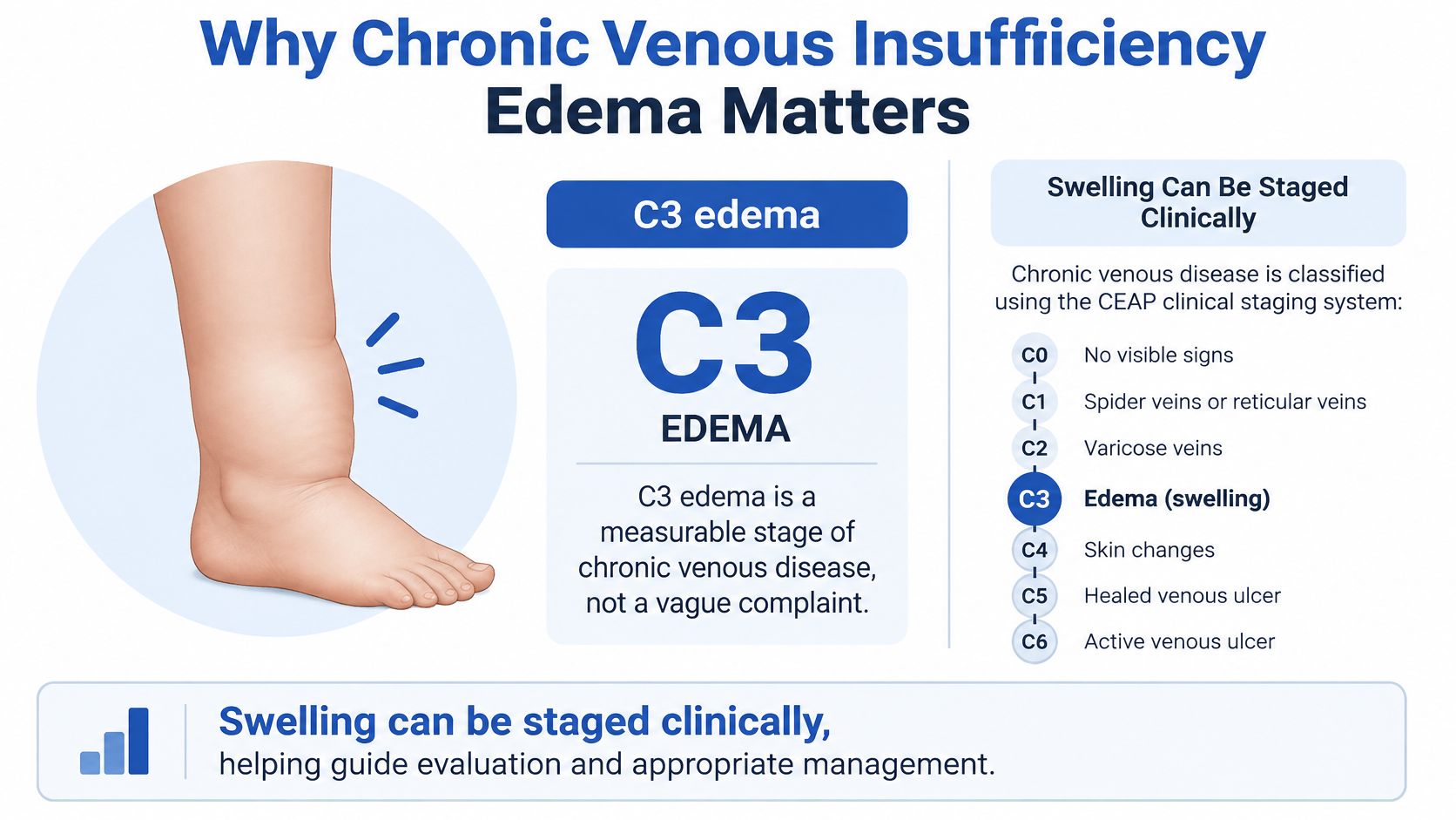
Edema isn't merely a cosmetic complaint or a vague feeling of puffiness. Clinicians can examine it, document its extent, and classify it as a defined manifestation of chronic venous disease.
The opening study illustrates an important distinction: swelling and ulceration aren't interchangeable measures of disease burden. Its findings describe the participants studied, not a universal estimate for every community or every person with swollen legs.
Why prevalence estimates differ
A study that counts visible varicose veins will answer a different question from one that counts edema, skin injury, or ulcers. Participants' ages, diagnostic methods, and the definition of venous insufficiency also affect the result.
That means apparently different prevalence estimates don't necessarily contradict each other. For an individual patient, the more useful question is whether examination and testing connect the swelling to a venous problem.
What swelling does and doesn't tell you
Swelling gives the clinician something concrete to investigate. It doesn't, by itself, establish which vein is affected, prove that a procedure is needed, or show that stronger compression would be safe.
Useful questions to bring to an appointment include:
- Is this swelling venous? Ask what examination findings and imaging support that conclusion.
- Could another condition contribute? Mention other medical conditions and bring a medication list.
- What should improve with treatment? Ask how the team will distinguish symptom relief from changes in skin health or circulation.
- What needs testing first? Clarify whether arterial assessment is necessary before choosing a stocking or bandage.
Treating swelling as a finding to explain, rather than something to squeeze away, changes the conversation. The clinician can build a plan around the cause, the condition of the skin, and the person's ability to use that plan consistently.
How the CEAP Classification Stages Venous Edema
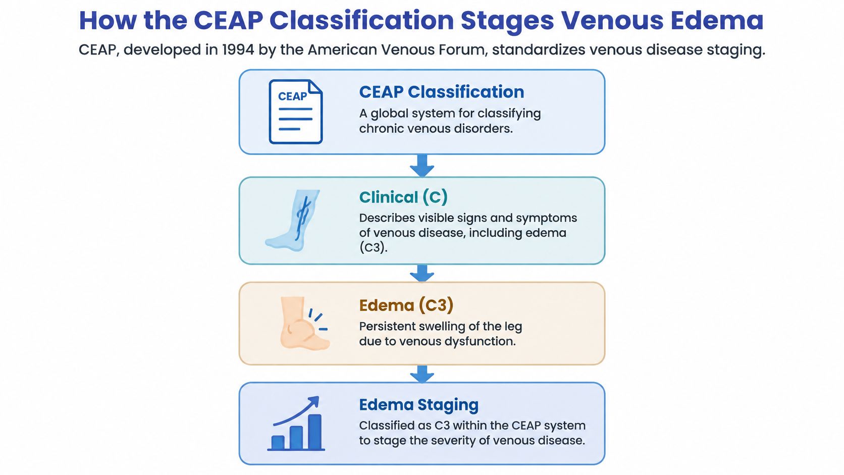
A report containing CEAP may look technical, but its purpose is practical: to describe venous disease consistently. The American Venous Forum developed the framework in 1994; its clinical categories range from C0 through C6, with edema classified as C3, as described in this review of venous disease classification.
The clinical categories distinguish no visible or palpable disease, small surface veins, varicose veins, edema, venous-related skin changes, healed ulcers, and active ulcers. Think of them as labeled findings, not a timetable predicting what will happen next.
Reading more than the clinical label
CEAP describes several dimensions of disease. The clinical label is only part of the assessment.
| Dimension | Plain-language meaning |
|---|---|
| Clinical manifestations | What the clinician can see or feel |
| Etiology | Why the venous problem developed |
| Anatomic distribution | Which veins are involved |
| Pathophysiology | Whether blood flows backward, encounters obstruction, or both |
This distinction matters when comparing people with similar-looking ankles. Someone with superficial vein reflux and someone with a history of deep-vein obstruction may share a swelling classification while needing different treatment discussions.
A stage isn't a prediction
Edema occupies a place between visible varicose veins and venous-related tissue injury in the clinical scale. That makes it useful for documenting disease, but it doesn't mean every swollen leg will develop an ulcer.
Nor does a classification establish the cause of every symptom. A patient can have documented venous disease and another contributor to swelling at the same time.
Practical interpretation: A CEAP label describes the venous findings. It doesn't replace an arterial assessment, an ultrasound explanation, or a review of other causes of edema.
Ask the clinician to translate the complete assessment, not just the letter-and-number shorthand. The most useful explanation connects the observed findings to the anatomy and the proposed treatment.
The Pressure Problem Behind Venous Swelling
Leg veins return blood toward the heart against gravity. The calf muscles provide a pumping action, while competent valves help prevent blood from falling backward between muscle contractions.
The pressure difference is substantial: foot-vein pressure while standing can reach approximately 80–90 mmHg, whereas effective calf pumping and competent valves normally reduce it to below 30 mmHg during walking, according to this review of chronic venous insufficiency mechanisms. With venous insufficiency, that walking-related pressure reduction is impaired.
Reflux and obstruction create different problems
Reflux means blood flows backward through valves that aren't closing effectively. Obstruction means blood meets resistance as it tries to leave the leg.
A useful analogy is a pump connected to a pipe. Leaky check valves allow backward flow; a narrowed outlet impedes forward flow. Either problem can keep downstream pressure high, even though the underlying defects differ.
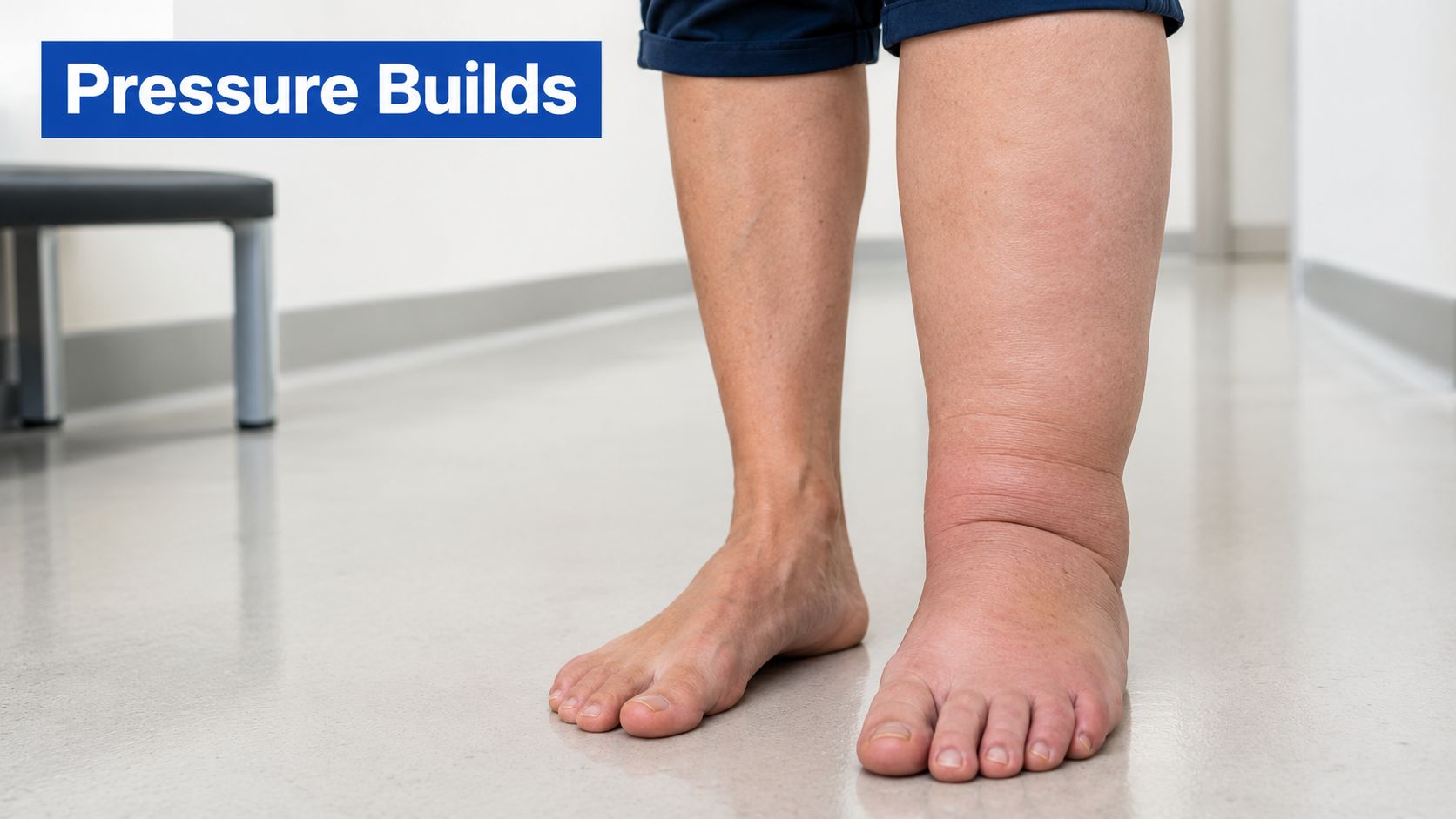
The tiny vessels supplying the tissues then face increased hydrostatic pressure. More fluid filters into the spaces between cells, which clinicians call the interstitial space.
Why the skin can change
Fluid movement depends partly on the pressure balance across small-vessel walls, often described using Starling forces. Chronic inflammation and activation of the vessel lining can also increase permeability, making those walls more leaky.
Over time, protein-rich fluid and inflammation can accompany tissue hardening, dark pigmentation, and lipodermatosclerosis, a firm change in the skin and underlying tissue. Advanced disease can include ulceration.
Several factors can add to the pressure problem:
- Reduced calf pumping: Immobility or restricted ankle movement limits the pumping mechanism.
- Greater venous load: Obesity and heart failure can add to the burden.
- Previous deep-vein thrombosis: A prior clot can leave obstruction or valve damage.
Swelling that becomes worse during prolonged standing and improves with elevation supports a venous explanation. It's a clue, however, not a substitute for establishing the cause.
Pitting Edema, Lymphedema, and Other Look-Alikes
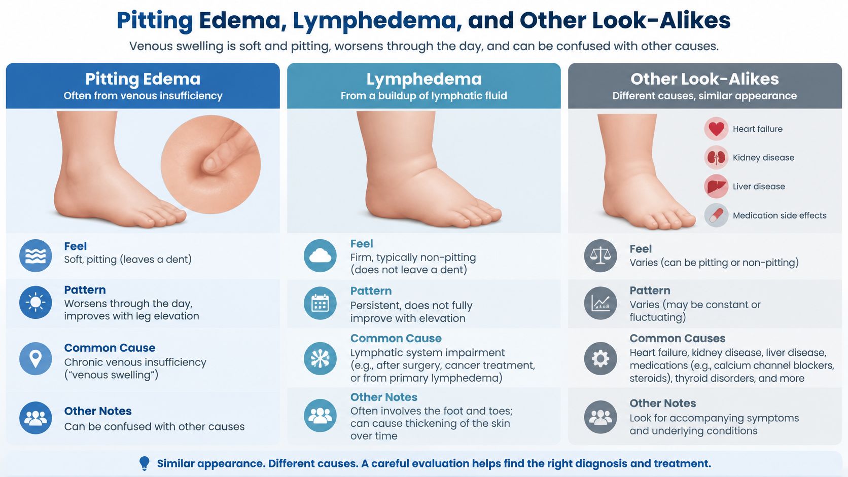
A dent left after pressing swollen skin describes pitting edema. It tells the clinician about the tissue's response to pressure, but it doesn't prove that venous insufficiency is the cause.
The pattern matters. Swelling associated with dependency, heaviness, varicose veins, or ankle staining fits a different clinical picture from swelling accompanied by broader signs of systemic illness.
Comparing possible contributors
| Possible contributor | What needs clarification |
|---|---|
| Venous disease | Does the pattern fit impaired venous return, and does imaging confirm it? |
| Lymphatic dysfunction | Is tissue-fluid drainage also impaired? |
| Cardiac or renal disease | Could a systemic condition contribute to fluid accumulation? |
| Medication effects | Did swelling change after a medicine was started or adjusted? |
| Post-thrombotic disease | Could an earlier clot have altered venous outflow? |
Lymphedema concerns the system that drains fluid from tissues, rather than the veins carrying blood back toward the heart. Persistent, firmer swelling can raise that possibility, but appearance alone isn't enough to classify it.
A dedicated lymphedema evaluation can help clarify whether lymphatic care belongs in the treatment plan. Venous and lymphatic problems aren't mutually exclusive.
Why arterial disease belongs in this discussion
Arterial disease isn't another name for venous insufficiency. The safety question is whether the leg receives enough blood before external pressure is applied.
Cold feet, absent pulses, pain with walking, diabetes, or concern about calcified arteries should prompt perfusion assessment. The clinician also needs to consider sensation, because someone who cannot reliably feel pressure-related discomfort needs a different monitoring approach.
Safety principle: Similar-looking swelling can require different treatment. Don't use appearance, a sock indentation, or another person's prescription to choose your compression.
Older adults and people with multiple medical conditions may have several contributors at once. A useful diagnosis identifies those contributors rather than forcing every swollen leg into a single category.
Assessing Edema Before Choosing Treatment
A good assessment begins with the history, not a stocking prescription. Tell the clinician when swelling began, whether it changes with standing or elevation, and whether you've had clots, wounds, reduced mobility, or changes in medication.
The examination should connect those details with skin findings, pulses, sensation, ankle movement, and the distribution of swelling. Bring previous ultrasound reports if they're available, but expect the clinician to interpret them alongside your current symptoms.
Checking arterial perfusion
The ankle-brachial index, or ABI, compares ankle and arm blood pressures. It helps guide whether standard compression, reduced compression, or further arterial evaluation is appropriate.
The Wound, Ostomy and Continence Nurses Society compression guidance uses these thresholds:
- ABI 0.8–1.3: Standard compression may be appropriate.
- ABI 0.5–0.8: Modified light compression may be appropriate under clinical supervision.
- ABI below 0.5 or above 1.3: Avoid compression while arranging arterial evaluation.
The shared boundary between the standard and modified ranges requires clinical interpretation. These categories aren't instructions for selecting a garment without an examination.
ABI also has limitations. Diabetes and arterial calcification can produce falsely high readings, so a high result isn't automatically reassuring when symptoms or examination findings suggest poor perfusion.
Making the ultrasound report understandable
Duplex ultrasound evaluates blood flow and venous anatomy. The clinician should assess relevant superficial veins, perforator veins connecting venous systems, and deep veins for reflux or obstruction.
Ask the report reader to separate the findings:
- Location: Which veins are abnormal?
- Mechanism: Is there reflux, obstruction, or both?
- Clinical relevance: Does that finding plausibly explain the swelling?
- Treatment implications: What would the proposed intervention address, and what might remain?
For example, finding superficial reflux doesn't automatically establish that it explains all the edema. The clinician still has to reconcile the scan with the examination and other medical conditions.
This assessment prevents an important mistake: escalating external pressure before confirming that the tissues can tolerate it. A garment should follow a circulation assessment, not stand in for one.
Compression, Procedures, and How to Choose
Compression applies external pressure to limit fluid moving into tissue and help blood return toward the heart. The right pressure depends on the clinical situation and whether the arteries can support it. The vascular guidelines on chronic venous disease describe approximately 20–30 mmHg at the ankle for uncomplicated symptomatic venous edema without significant arterial insufficiency. For venous ulceration, they describe commonly 30–40 mmHg, or at least 40 mmHg with multilayer or inelastic systems.
Compression Pressure Targets by Clinical Situation
| Clinical situation | Typical ankle pressure | Key caution |
|---|---|---|
| Uncomplicated venous edema | Approximately 20–30 mmHg | Significant arterial insufficiency must be excluded |
| Venous ulceration | Commonly 30–40 mmHg, or at least 40 mmHg with multilayer or inelastic systems | Requires wound assessment and an appropriate monitoring plan |
| Mixed arterial and venous disease | No routine target; pressure must be reduced or otherwise modified to match arterial perfusion | Avoid routine high-pressure treatment; a clinician should set the pressure |
| Uncertain perfusion | No pressure selected until evaluated | Establish safety first |
A compression therapy consultation can help match garment length and size to skin condition, hand strength, mobility, and available help at home. Applying compression in the morning, before swelling builds while the legs are down, can improve fit.
Supportive care and procedures serve different purposes
For someone using a compatible mobility device, positioning equipment may help with a clinician-approved elevation plan. Patients can find elevating leg rests for Jazzy, then confirm compatibility and safe positioning with their equipment team.
Procedures treat selected abnormal veins. Depending on vein anatomy, options include thermal ablation, adhesive closure, mechanochemical treatment, ultrasound-guided foam sclerotherapy, or removal of selected superficial varicose veins. For active venous ulcers, appropriately selected early treatment of relevant superficial reflux can speed healing alongside compression. A swollen ankle alone does not mean a procedure is needed.
Pneumatic compression may be considered when swelling persists despite standard compression or stockings are difficult to apply. The plan needs to account for cardiac, renal, infectious, and arterial concerns, since these can change whether compression is suitable.
Higher pressure is not automatically better: a garment that the skin cannot tolerate or the arteries cannot support does more harm than good. A useful comparison is a shoe that fits: added support only helps when it suits the person wearing it.
Some people need both a garment and a procedure; others need heart, kidney, or arterial evaluation first, because compression or ablation will not address those causes. Venous edema is only one possible source of leg swelling, so treatment should follow the cause rather than the appearance of the ankle alone.
Why Swelling Can Persist After Vein Treatment
Persistent edema after vein treatment doesn't automatically mean the procedure failed. It does mean the remaining swelling deserves an explanation rather than an assumption that another procedure or tighter garment will solve it.
Phlebolymphedema describes combined venous and lymphatic dysfunction. Chronic venous hypertension can overload lymphatic drainage, leaving a tissue-fluid problem that may persist even after a relevant refluxing vein has been treated.
Treating the drainage problem as well
A 2025 randomized clinical trial found that four weeks of complex decongestive therapy combining manual lymphatic drainage, intermittent pneumatic therapy, and bilayer bandaging improved pain, heaviness, venous flow velocity, venous severity scores, and quality of life, as reported in the trial of complex decongestive therapy for chronic venous insufficiency. Those findings support considering lymphatic-directed care in an appropriate patient, not promising that every person's swelling will disappear.
Nor should medication be treated as a guaranteed substitute for addressing the underlying contributors. A clinician needs to evaluate the likely benefit and suitability of any proposed medicine for the individual patient.
Reassessing what remains
Follow-up should distinguish among several possibilities rather than grouping them together as residual edema:
- Remaining venous disease: Review whether reflux or outflow obstruction still contributes.
- Lymphatic involvement: Assess whether a coordinated decongestive program is appropriate.
- Other contributors: Revisit medications, obesity-related swelling, cardiac or renal disease, and infection.
- Practical treatment barriers: Check whether the prescribed garment fits and whether it can be applied.
The timing of reassessment should follow the treatment plan and the person's symptoms. Improvement can be gradual, and the clinician should explain what changes would be reassuring and what would trigger further investigation.
Skin leakage, dermatitis, difficulty applying compression, or recurrent wounds make coordinated vein-and-wound care especially useful. The team can reassess the circulation while adapting skin protection and edema management.
A realistic treatment goal may be less swelling, healthier skin, improved comfort, and easier daily activity rather than an immediate return to an entirely unswollen leg. Agreeing on those goals makes follow-up more meaningful.
Monitoring Swelling and Preventing Complications
Monitoring works best when you compare like with like. Ask your care team where to measure ankle and calf circumference, when to record weight, and which changes should prompt a call.
A brief record can help distinguish a changing pattern from an isolated difficult day. It should support clinical review, not become a reason to adjust compression pressure yourself.
What to track
- Swelling and fit: Record changes in circumference, shoe tightness, and how easily the prescribed garment goes on.
- Skin condition: Note new staining, hardening, itching, dermatitis, leakage, or broken skin.
- Comfort and sensation: Report numbness, pressure pain, or worsening discomfort with walking.
- Wounds: Seek assessment for a new opening or a wound that isn't healing; venous leg ulcer care should address circulation alongside the wound.
When to seek urgent help
Sudden one-sided swelling needs prompt medical assessment rather than an increase in compression. Chest pain or new breathlessness warrants emergency evaluation.
Fever, spreading redness, new wounds, or a cold, painful or numb foot also require prompt attention. Don't assume these are ordinary fluctuations in chronic edema.
Routine reassessment is part of treatment, not evidence that treatment has failed. Persistent or recurrent swelling, especially with skin changes, warrants a venous evaluation and an arterial assessment appropriate to the compression decision.
Vein and Wound Institute offers clinical examination, onsite duplex ultrasound, venous treatment, and coordinated wound care for patients with persistent leg swelling and related skin injury. Visit Vein and Wound Institute to arrange an evaluation and discuss a compression and follow-up plan based on your circulation, medical conditions, and daily needs.