
Most advice about circulation problems starts with blocked arteries and ends with a pulse check. That leaves out the part that feeds a wound, the microcirculation, where oxygen, nutrients, and waste exchange happens at the capillary level. If the small vessels aren't delivering enough oxygen, tissue can fail even when a larger artery is open.
Table of Contents
- Why Poor Tissue Perfusion Is Often Misunderstood
- Vascular and Non-Vascular Causes of Reduced Perfusion
- Diagnostic Testing Beyond the Ankle-Brachial Index
- Understanding Transcutaneous Oxygen Pressure for Wound Care
- How Poor Perfusion Impairs Wound Healing
- Treatment Principles for Improving Tissue Perfusion
- When to Seek Immediate Evaluation for Non-Healing Wounds
Why Poor Tissue Perfusion Is Often Misunderstood
A strong pulse can feel reassuring, but it does not prove that skin, muscle, or a wound bed is getting enough oxygen. Poor tissue perfusion is a tissue-level problem that a large-artery exam can miss. A leg may look well supplied on a quick check and still struggle to heal.
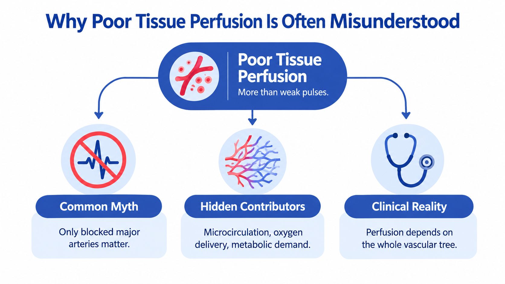
Why the old shortcut falls short
The history of microcirculation makes the point clearly. Marcello Malpighi identified capillaries as the bridge between arteries and veins, Antoni van Leeuwenhoek confirmed capillary perfusion in 1688, and August Krogh later earned the 1920 Nobel Prize for work on capillary motor regulation AHA review. Those milestones show that tissue viability depends on what happens in the smallest vessels, not only on whether a big vessel is open.
Practical rule: If the wound bed is starving for oxygen, a palpable pulse does not solve the problem.
Impaired microvascular perfusion is common in people with hypertension, diabetes, obesity, and dyslipidemia. It can also appear early in hypertension and diabetes, long before the problem feels advanced. That helps explain why some ischemic, diabetic, and postoperative wounds stall instead of moving through normal healing.
What this means at the bedside
A wound needs enough oxygen for collagen formation, immune defense, epithelial migration, and resistance to infection. If delivery is low, the tissue cannot complete those steps efficiently. The result is often a wound that looks inflamed, sluggish, or repeatedly open, even when the pulse seems normal.
Vascular and Non-Vascular Causes of Reduced Perfusion
Peripheral artery disease is one of the clearest examples of chronic poor circulation. The Global Burden of Disease 2019 analysis estimated 113 million adults aged 40 years and older had PAD worldwide in 2019, with prevalence rising sharply in older age groups Lancet Global Health00355-8/fulltext). That is one reason older adults and people with cardiovascular risk factors deserve a circulation-focused look when a wound does not close.
Arterial disease is only part of the picture
PAD can stay quiet for a long time. When it progresses, oxygen delivery to the lower extremities falls, and the tissue may respond with exertional pain, rest pain, ulcers, tissue death, and a higher risk of amputation Lancet Global Health. Ischemic ulcers often appear on the toes or heel, especially after minor trauma, so a small injury can become a major wound when flow is limited peripheral arterial disease and poor circulation.
A useful way to sort reduced perfusion is into two broad groups:
- Vascular causes: arterial obstruction, arterial stiffness, and microvascular dysfunction.
- Non-vascular contributors: venous congestion, edema, lymphedema, neuropathy, infection, pressure, and poor metabolic control.
These non-vascular factors matter because they can reduce tissue oxygen delivery even when the main artery is not fully blocked. Venous pooling and swelling increase the distance oxygen must travel, like trying to carry water through a hose that is partly pinched and surrounded by standing water. Neuropathy hides pain, so pressure and friction continue unchecked. Infection raises local demand while also disrupting delivery. Poor metabolic control weakens the tissue response on several fronts at once. The result can look like “poor circulation” even when more than one mechanism is involved.
Why overlap creates confusion
A person with diabetes may have arterial disease, neuropathy, and edema at the same time. Another patient may have venous reflux plus a smaller arterial problem, and the wound still will not behave like a simple ulcer.
That overlap is why a single label rarely tells the full story. The bedside question is not only whether an artery is open, but whether oxygen is reaching the wound bed in enough quantity to support repair. A pulse can be present and perfusion can still be inadequate at the tissue level.
A wound often tells you more than the pulse exam does.
Diagnostic Testing Beyond the Ankle-Brachial Index
The ankle-brachial index is a useful starting point, but it does not answer every question about wound perfusion. An ABI of 0.71 to 0.90 suggests mild PAD, 0.41 to 0.70 moderate disease, and ≤0.40 severe disease PMC review. An ABI above 1.31 can look normal while calcified arteries stay stiff and resist compression, so the result may be falsely reassuring PMC review. That problem is common in diabetes, renal disease, and medial arterial calcification.
Match the test to the question
A practical workup starts with the clinical question. If you want to know whether there is obvious large-vessel disease, ABI can help. If you want to know whether oxygen is reaching the wound bed in a way that supports repair, toe pressure, toe-brachial index, Doppler waveforms, skin perfusion pressure, and TcPO₂ are more informative. The diagnostic laboratory evaluation should be chosen to answer what the wound needs, not just what the limb looks like.
A simple way to organize the workup is this:
- Start with ABI and exam. This gives a broad view of arterial disease.
- Escalate when ABI may mislead. Diabetes and calcification can distort the result.
- Use toe-based or oxygen-based measures. These better reflect tissue-level perfusion.
- Act on the wound. The test should answer whether the wound can plausibly heal.
A toe-brachial index below 0.70 is abnormal, and toe pressure below about 30 to 50 mmHg is associated with impaired healing potential PMC review. These values do not prove that healing will fail, but they do suggest the tissue is working with limited oxygen reserve.
Why that hierarchy matters in real life
A patient can have a wound, a palpable pulse, and a borderline ABI, yet still have tissue that is too underfed to heal well. In that setting, a normal-seeming limb exam can hide a problem at the skin level. The better question is whether oxygen delivery is enough where repair has to happen.
Understanding Transcutaneous Oxygen Pressure for Wound Care
TcPO₂ measures how much oxygen is available in skin near an ulcer at one specific site. That local reading is often more useful for wound decisions than a test that only reflects larger vessels. Values above roughly 55 to 70 mmHg are generally compatible with adequate oxygenation, while values below 40 mmHg suggest impaired healing potential and values below 30 mmHg fit severe ischemia or critical compromise CCJM.
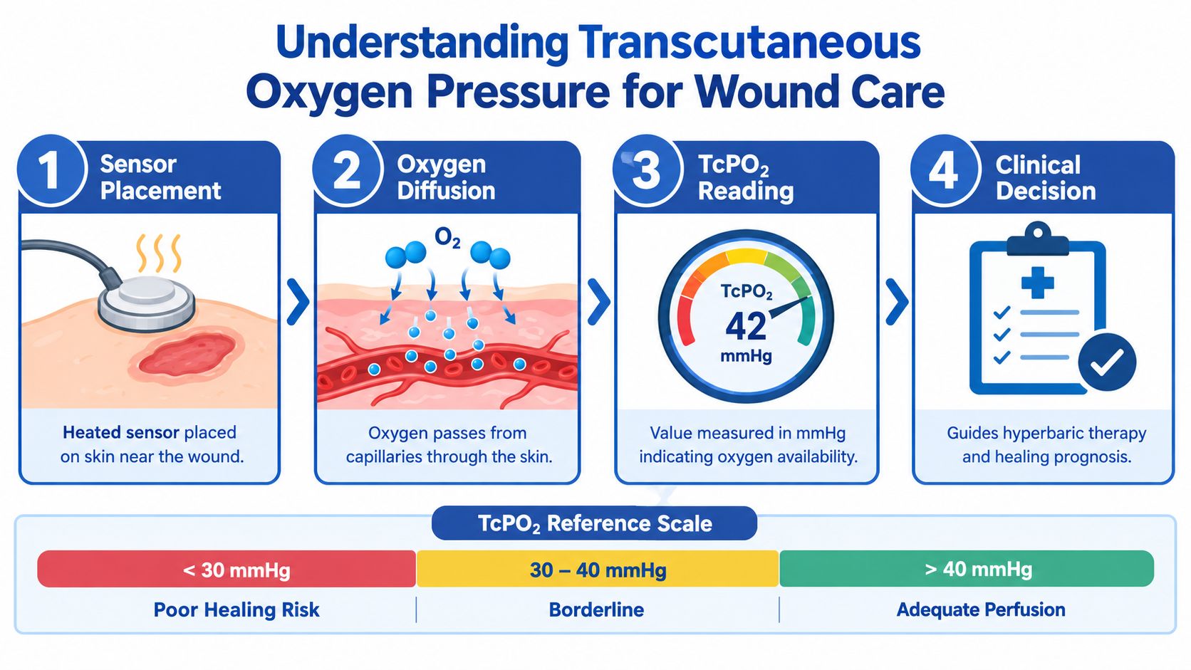
How the test works
The sensor is heated and placed on intact skin near the wound, usually at a standardized location. Oxygen then diffuses through the skin, and the monitor estimates how much is available in that area. The number you get is a local oxygen readout for that specific skin site.
Clinical insight: A rising TcPO₂ after revascularization is reassuring because it shows oxygen delivery has improved where healing has to occur.
That matters when you want to know whether treatment changed the biology around the wound. Persistently low or falling values should prompt reassessment for arterial disease, edema, infection, pressure, anemia, or hypoxemia. The number does not replace judgment, but it sharpens it CCJM.
How to interpret the result
A small diabetic foot ulcer with a TcPO₂ of 24 mmHg was associated with healing prediction in one clinical cohort, with reported sensitivity of 0.89. That does not mean every wound behaves the same way, and it does not guarantee outcome. It does show why oxygen-based testing can outperform a simple “looks okay” assessment CCJM.
The most useful TcPO₂ studies are standardized. The sensor should sit on intact periwound skin, the location should be documented, the patient position should be noted, and the test should be repeated after an intervention or when the wound changes CCJM. That creates a trend, and trends are often more valuable than a single value.
How Poor Perfusion Impairs Wound Healing
Healing slows when oxygen delivery stays low. Sustained hypoxia weakens angiogenesis, collagen deposition, fibroblast activity, and epithelialization, while inflammation lasts longer than it should. The wound stays in a repair mode that never fully completes.
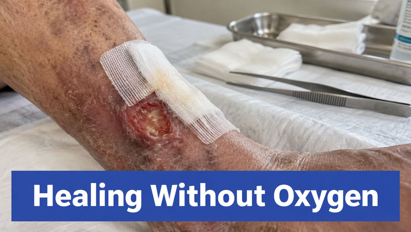
Why stalled healing keeps repeating
A wound needs collagen for strength, fibroblasts for matrix building, and new vessels to support repair. If oxygen supply is poor, each step runs slowly, like a worksite waiting on deliveries that never arrive. The wound can then move through drainage, partial closure, and breakdown without becoming stable.
That pattern also helps explain why infection risk rises and why dressings can seem less effective than expected. In people with diabetes, diabetes care conditions matter because glucose control, infection control, and oxygen delivery all shape the same healing environment.
Why revascularization alone may not close the wound
A reopened vessel helps only if tissue-level oxygen delivery improves as well. If microvascular dysfunction remains, the skin can still behave as though supply is limited. That is why a technically successful vascular procedure does not always translate into closure.
Arterial leg ulcers show this clearly, since flow in a larger artery is only part of the story. The wound bed still has to receive enough oxygen at the capillary level for repair to keep moving.
Treatment Principles for Improving Tissue Perfusion
Treatment works best when it improves perfusion at the tissue level and supports the wound bed at the same time. That means reducing obstruction where it exists, improving oxygen delivery where it is weak, and protecting tissue while repair catches up. A coordinated plan may include vascular evaluation, wound debridement, compression when appropriate, pressure relief, infection control, and follow-up that tracks objective change.
Build the plan around perfusion, not assumptions
The first step is to identify what is limiting flow. If venous reflux is driving swelling and pressure, treating that helps the tissue environment. If arterial disease is reducing inflow, the plan may shift toward imaging and possible intervention. When the perfusion problem is mixed, both sides matter.
A wound-focused clinic can help when diagnostics and treatment sit in the same place, because the patient does not get bounced between separate systems. Vein and Wound Institute shows that approach in practice by pairing vascular testing, wound care, and longitudinal monitoring in one setting. The point of that model is straightforward, to track perfusion, wound dimensions, and response together instead of treating them as separate problems.
Support healing while the circulation is corrected
Compression can help selected patients with venous disease, but it has to be matched to arterial status first. Bioengineered skin substitutes and pressure wound therapy may help the wound bed, but they do not replace oxygen delivery. They work best when the underlying circulation issue is being managed at the same time.
Treatment should measurably improve the wound bed, not just its appearance.
For patients who need structured chronic-care support, Core Nursing Solutions chronic care can help reinforce follow-up, medication adherence, and monitoring between clinic visits. Consistency matters. Poor perfusion improves when the plan is coordinated, reassessment is scheduled, and the patient knows which changes need prompt attention.
When to Seek Immediate Evaluation for Non-Healing Wounds
A wound can look quiet and still be poorly perfused. A person with diabetes may say, “It doesn't hurt, so I thought it was fine.” A person with numb feet may only notice that a small ulcer has stopped changing. Those stories are common, and they are risky, because absence of pain doesn't prove good blood flow in diabetic or neuropathic feet IWGDF guidance. No single symptom reliably predicts healing.
Red flags that should not wait
Seek prompt evaluation if a wound is getting larger, developing new discoloration, showing tissue necrosis, or sitting on a cool foot. Rest pain, diminished Doppler signals, or obvious failure to progress are also warning signs, and the WIfI framework treats these findings as markers that deserve urgent attention.
A few patterns should push the workup forward:
- Cooler skin, worse color, or dead tissue: act quickly.
- Wound dimensions increasing instead of shrinking: reassess perfusion.
- Acceptable-looking imaging but poor wound progress: ask for functional testing.
That last point matters. An angiogram or Doppler study can look acceptable while the wound still lacks enough oxygen at the tissue level. In that situation, toe pressure, TcPO₂, or another functional measure is often more useful than a reassuring snapshot.
If the wound has stalled, the operative question is whether the tissue is recovering, and functional testing answers it.
If you're dealing with a wound that hasn't improved, or a leg that keeps swelling, changing color, or feeling colder than it should, the next step is a focused circulation workup. Vein and Wound Institute provides vascular testing and wound care in one setting, which can help connect perfusion findings to a real treatment plan.