
A reticular vein is a small, usually blue-green superficial vein that measures about 1 to 3 mm in diameter and sits between the skin and the deeper fascia. It's distinct from a spider vein, which is usually smaller, and from a varicose vein, which is larger and more prominent when you're standing.
You may have noticed a faint blue line on your thigh or calf and wondered whether it means something serious. In many cases, the answer depends less on how it looks and more on what else is happening in the leg, which is why the same visible vein can be a cosmetic finding for one person and a clue to deeper venous reflux for another.
Table of Contents
- What Is a Reticular Vein
- Reticular Veins vs Spider Veins and Varicose Veins
- Understanding the Anatomy and Blood Flow Connection
- When to Seek Evaluation and Ultrasound Testing
- Treatment Options and Recent Evidence
- Making an Informed Treatment Decision
- Next Steps for Managing Reticular Veins
What Is a Reticular Vein
A reticular vein is a dilated superficial vein that usually looks bluish and slightly twisted under the skin. It most often appears on the lower limbs, where pressure, standing, and venous valve stress tend to show up first.
For many patients, the first clue is simple. They see a faint blue network or a single wavy line and assume it's just a cosmetic issue. That assumption can be right, but not always.
A reticular vein typically measures about 1 to 3 mm across, although some clinical classifications extend the upper limit to 4 mm (clinical review). That size matters because it places reticular veins between spider veins and larger varicose veins, both in appearance and in clinical meaning.
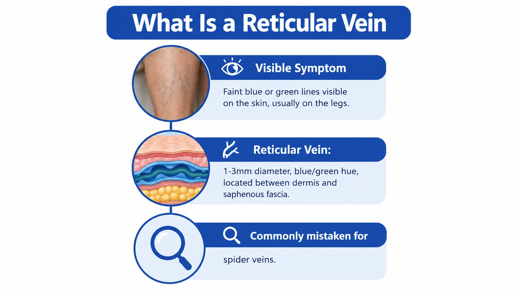
A visible vein is a finding, not a diagnosis by itself.
The part many people miss is location. Reticular veins sit in the subdermal tissue and form part of a network that can drain into larger superficial veins, so the visible line you see on the surface may be connected to a broader venous pattern. That is why understanding what is a reticular vein is the first step before deciding whether to observe it, evaluate it, or treat it.
Reticular Veins vs Spider Veins and Varicose Veins
A patient may notice a faint blue web on the leg, wonder whether it is harmless, and then notice a thicker rope-like vein nearby. That comparison is the quickest way to separate the three common surface vein types. Spider veins are the finest, reticular veins are intermediate, and varicose veins are larger, more prominent veins that are easier to see and sometimes to feel when standing (comparative clinical review).
Size and depth tell the story
Spider veins, also called telangiectasias, are typically less than 1 mm and lie within the skin itself. Reticular veins are usually 1 to 3 mm, sometimes described up to 4 mm, and sit deeper, between the dermis and the saphenous fascia (anatomical review). Varicose veins are generally 3 mm or larger and visibly tortuous or bulging when the patient is standing.
That difference is more than cosmetic detail. It helps a clinician judge whether the visible vein is likely an isolated surface finding or part of a broader reflux pattern. A visible vein is a finding, not a diagnosis, and context determines what comes next.
| Vein Type | Diameter | Depth | Appearance | CEAP Class |
|---|---|---|---|---|
| Spider Veins | Under 1 mm | Intradermal | Fine red, blue, or purple webbing | C1 |
| Reticular Veins | About 1 to 3 mm, sometimes up to 4 mm | Between dermis and saphenous fascia | Blue-green, slightly tortuous lines | C1 |
| Varicose Veins | Generally 3 mm or larger | Superficial, more prominent | Bulging, rope-like, tortuous veins | Higher than C1 |
All three fall under superficial venous disease, but they do not mean the same thing anatomically. A reticular vein can remain a cosmetic finding, or it can act as a feeder channel for spider veins and reflect underlying reflux. That is why appearance alone cannot answer the whole question.
Learn how spider veins and varicose veins differ in practice
Understanding the Anatomy and Blood Flow Connection
Reticular veins are part of a horizontal superficial network between the dermis and the saphenous fascia. They collect blood from the skin and subcutaneous tissues, then communicate with larger superficial veins that return blood toward the heart (anatomical review).
Why valves matter
In a healthy leg, one-way valves keep blood moving in the right direction. The calf-muscle pump helps push blood upward when you walk, and perforator veins normally direct flow from the superficial system into the deep system. When a perforator becomes incompetent, pressure from the deep system can push backward into the superficial network (hemodynamic review).
That backward pressure can widen small surface veins, increase tortuosity, and create reticular or spider-vein patterns. It's a bit like a plumbing branch with a faulty valve, pressure builds where it shouldn't, and the smaller lines start showing strain.
Reticular veins are important because they can sit at either end of the spectrum. Some are isolated and mainly cosmetic. Others are visible markers of altered venous hemodynamics, especially when they cluster with spider veins or come with aching, heaviness, swelling, or skin change.
See how ultrasound helps identify the source of reflux
What a careful exam looks for
A focused exam doesn't stop at the vein itself. It should document where the vein is, whether there are symptoms, and whether edema, skin changes, or other varicosities are present. That context helps separate a small cosmetic vein from a pattern that deserves deeper mapping.
When to Seek Evaluation and Ultrasound Testing
A visible reticular vein does not automatically mean imaging is needed. If the vein is isolated, painless, and not accompanied by swelling or skin change, watching it over time can be reasonable. The central question is whether it is acting alone or reflecting a broader venous problem.
Signs that make evaluation more important
Evaluation becomes more important when the vein appears with:
- Aching or heaviness: These symptoms can point to reflux elsewhere in the venous system.
- Swelling: Edema shifts the concern beyond a surface cosmetic finding.
- Skin changes: Color change, irritation, thickening, or a wound suggest more advanced venous disease.
- Recurrent visible veins: Veins that return after prior treatment can mean the underlying source has not been addressed.
- Associated varicosities: Larger nearby veins make reflux more likely.
A clinician uses duplex ultrasound to map the great and small saphenous veins, tributaries, perforators, and deep veins, so the source of reflux can be identified. That matters because the visible reticular vein may be only the surface sign of a deeper pressure problem. Small intradermal vessels may not be fully evaluated on standard scanning, but the connected venous pathways often can be (Vein and Wound Institute's ultrasound guide).
If symptoms are part of the picture, the goal is not to chase the visible line. It is to find out whether that line is pointing to something deeper.
A practical rule helps. A quiet cosmetic vein may only need monitoring. A vein that comes with discomfort, swelling, skin change, or a pattern of larger veins deserves ultrasound mapping.
Treatment Options and Recent Evidence
Reticular veins are often treated with sclerotherapy because their usual 1 to 3 mm size fits injection-based treatment well. The aim is to close the visible vein, soften its appearance, and, when needed, treat feeder veins that support spider-vein clusters. For a plain-language overview of the procedure, see how sclerotherapy works for visible veins.
What patients should expect
Treatment can improve what you see, but it does not erase the tendency to form visible veins. Some people need more than one session. Some are left with nearby veins that were too small, too deep, or too spread out to clear in one visit. Pigmentation changes and matting can also happen, so the goal should be improvement, not perfection.
A randomized trial in 41 patients compared polidocanol sclerotherapy alone with transdermal radiofrequency plus polidocanol. After one session, both approaches reduced treated vessel length, with average reductions of 30.7% for sclerotherapy alone and 33% for the combined approach, with no statistically significant difference between groups (trial report). The same trial also reported improvements in quality of life and pain reduction.
That finding matters because newer energy-based add-ons are sometimes assumed to work better than injection treatment. In this study, they did not clearly do so. For patients, the practical lesson is simple, match the treatment to the anatomy and the goal, rather than assuming the newest option is automatically better.
How to think about choice
- If the vein is cosmetic and small: sclerotherapy is often the practical starting point.
- If symptoms suggest reflux: treatment planning should include evaluation of the source, not just the surface vein.
- If the pattern is complex: the visible reticular vein may be only one part of the problem, so treatment may need to be staged.
Making an Informed Treatment Decision
A patient may ask, “Is this just a cosmetic vein, or does it point to a reflux problem?” That question gets to the heart of the decision. The right choice depends on vein size, depth, anatomy, symptoms, and ultrasound findings when imaging is warranted. Two people can have veins that look similar on the surface and still need different care.
Cosmetic treatment is reasonable when a reticular vein is isolated and not causing symptoms. Evaluation matters more when aching, heaviness, swelling, recurrence, or skin change suggests the vein may be part of a larger venous pattern. Treat the vein you can see, but don't ignore the circuit it may belong to.
A reticular vein can be treated for appearance, or it can be studied as a possible marker of venous reflux. Those are separate decisions. One is about how the vein looks, the other is about whether blood flow is being directed the wrong way.
Patients should also know what treatment can and cannot do. Veins can return, and new ones can appear, because the tendency to form them may still be present. That does not mean care was a failure. It means planning should be realistic, and some people need staged treatment.
Ask three direct questions in the consult: what is this vein connected to, is imaging needed, and is the goal appearance, symptom relief, or both? Those answers help separate a simple cosmetic procedure from a broader venous treatment plan.
Next Steps for Managing Reticular Veins
Start with observation if the vein is small, stable, and not causing symptoms. Take note of whether it's changing, whether more veins are appearing, and whether the leg feels heavy, tired, or swollen by the end of the day. Those details help a clinician decide if the vein is merely visible or clinically meaningful.
If you notice aching, heaviness, swelling, skin color changes, or recurrent veins, schedule an evaluation. Bring a history of previous vein treatment, leg wounds, clotting issues, or long-standing swelling if any of those apply. A careful exam and, when needed, duplex ultrasound can sort out whether the visible vein is isolated or part of a deeper reflux pattern.
The most useful care comes from a specialist who can assess both venous disease and skin healing concerns when needed. In that setting, treatment can be matched to the actual problem instead of the surface appearance alone. For some people, that means reassurance and monitoring. For others, it means minimally invasive treatment with a clear plan for follow-up.
If you're trying to decide whether a reticular vein on your leg is harmless or worth a closer look, the team at Vein and Wound Institute evaluates visible veins, checks for reflux when it matters, and helps patients choose between observation and minimally invasive treatment based on their anatomy and symptoms. Visit their site to review care options and arrange an evaluation if your veins are changing, uncomfortable, or accompanied by swelling or skin changes.