
You've had an ultrasound because one leg suddenly became swollen, or because aching, visible veins, or ankle swelling has been getting harder to ignore. The report says there's no deep-vein thrombosis, yet your symptoms continue. That situation is common because a DVT-focused ultrasound and a complete venous reflux map are not necessarily the same examination.
Vein mapping of legs uses duplex ultrasound to show the anatomy of the veins and how blood moves through them. A thorough study can identify clots or obstruction, faulty valves, backward flow, and the pathways connecting deeper veins with surface veins. That information helps a vascular specialist choose treatment based on the actual source of the problem rather than on the most visible vein.
Table of Contents
- Why Your Leg Veins Need a Road Map
- What Duplex Ultrasound Actually Measures
- How the Mapping Examination Is Performed
- Reading Reflux Results and Their Treatment Implications
- When a Normal Scan May Still Miss Venous Disease
- Mapping for Wounds, Diabetes, and Mixed Disease
Why Your Leg Veins Need a Road Map
Swollen ankles, aching legs, heaviness, visible varicose veins, and a wound that won't heal can arise from different circulation problems. A clinician needs to determine whether blood is moving normally toward the heart, pooling because valves aren't closing properly, or meeting resistance because a vein is obstructed.
Venous mapping provides that road map. It examines the deep veins, superficial veins near the skin, and perforating veins that connect the two systems. The study can document whether a vein is open, whether it compresses normally, where reflux begins, and how far that abnormal flow travels.
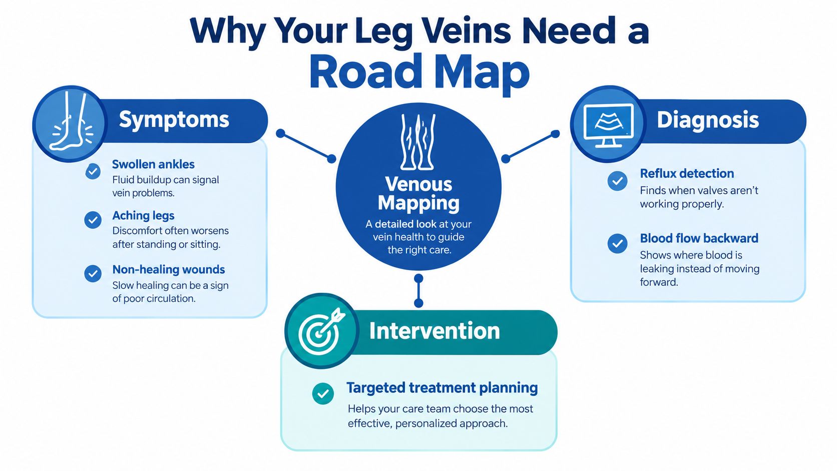
From symptoms to a treatment target
Think of the visible varicose vein as a branch rather than the trunk. Treating only that branch may leave the upstream source of reflux untouched. A map helps the care team trace the abnormal pathway and decide whether the important finding is in a saphenous vein, a tributary, a perforator, or the deep system.
This matters for everyday symptoms and for advanced disease. In a population study summarized in medical literature, 84% of participants had some form of chronic venous disease beyond the absence-of-disease category, and 41.4% had early clinical stages marked by telangiectasias or varicose veins (medical literature on chronic venous disease and leg ulcers). Those figures don't mean every swollen leg has venous disease, but they show why persistent symptoms deserve more than a surface inspection.
Venous leg ulcers represent the advanced end of this spectrum and affect approximately 1% of adults (evidence on chronic venous disease and ulcer burden). Mapping can connect the wound to reflux or obstruction, establish a baseline before treatment, and help clinicians monitor whether the circulation problem remains active.
Practical rule: A scan should answer the clinical question your symptoms raise. “No DVT” is reassuring for clot evaluation, but it may not answer whether faulty valves are causing chronic reflux.
For background on how long-term vein disease develops and affects the legs, see this explanation of chronic venous disease.
What Duplex Ultrasound Actually Measures
Duplex ultrasound combines structural imaging with blood-flow assessment. It is not just a photograph of veins. A technically useful examination brings together four related tools, each answering a different question.
The four parts of the examination
-
B-mode grayscale imaging shows the vein's shape, walls, surrounding tissue, and internal contents. It helps the examiner follow the vessel and look for material that could represent thrombus or another obstruction.
-
Compression testing checks whether the vein collapses under gentle pressure from the probe. A healthy, open vein should compress in the expected segments. Failure to compress can raise concern for thrombus, although the finding must be interpreted with the rest of the examination.
-
Color Doppler overlays moving color on the grayscale image. It helps the examiner see whether blood is flowing, identify flow direction, and follow connections between vessels. Color is useful for orientation, but a persistent color pattern doesn't reliably tell the examiner how long flow has reversed.
-
Spectral Doppler converts flow into a waveform. The examiner can use calipers on that trace to measure the duration of backward flow, or reflux, after a maneuver that changes venous pressure. This measurement helps identify whether reflux is brief, sustained, and clinically relevant in a particular segment.
The combination matters because a static color image can't quantify reflux duration or reliably identify its source. The map should show anatomy and function together, including the deep and superficial systems, perforators, obstruction, respiratory phasicity, augmentation response, and the route of abnormal flow (lower-extremity venous mapping guidance).
Why the report uses unfamiliar abbreviations
You may see GSV, meaning great saphenous vein, or SSV, meaning small saphenous vein. An accessory vein is an additional channel that can contribute to drainage or reflux. A perforator passes between superficial and deep systems, so the examiner may document whether it carries abnormal flow toward the skin or deeper tissues.
The result isn't just a list of vessels. It should explain where the abnormal flow starts, which branches it reaches, and whether the pattern supports a treatment directed at an axial vein or a smaller tributary.
If you need to plan the examination financially or compare what different ultrasound services include, a general guide to ultrasound costs in Dubai may help explain why pricing depends on the type and extent of imaging. For related information about diagnostic evaluation and treatment planning, see ultrasound diagnosis for varicose and spider veins.
How the Mapping Examination Is Performed
A complete study follows a sequence. The technologist isn't just moving a probe over the most visible veins. Position, pressure, flow maneuvers, and documentation all affect what the examination can reveal.
First, the examiner checks patency
You may begin lying down, or supine. This position supports assessment of the deep veins for openness and possible thrombus. The technologist applies ultrasound gel and moves the transducer along the groin, thigh, knee, calf, and ankle regions as clinically appropriate.
Compression testing occurs during this part of the examination. The examiner also observes respiratory phasicity, which describes how venous flow changes with breathing, and augmentation, which describes the response when flow is increased through a gentle maneuver.
Then, the leg becomes dependent
Reflux testing works differently from a clot check. The limb should be dependent, meaning the patient is standing, sitting, or positioned in reverse Trendelenburg with the head raised. Guidance commonly recommends a dependent position of at least 15 degrees for reflux assessment because gravity increases venous filling and can expose valve incompetence (lower-extremity venous mapping guidance).
Standing isn't a test of endurance. If standing isn't safe or practical, the technologist may use another dependent position. You may be asked to bear down or perform a calf maneuver while the examiner records the resulting Doppler waveform.
Finally, the technologist traces the pathway
The examination should document the deep veins, the GSV and SSV systems, relevant tributaries, and perforators. It can record vein diameter at standardized locations, reflux duration, the distal extent of reflux, and whether the pathway is continuous or limited to a short segment.
When a unilateral study is appropriate, the common-femoral spectral waveform may be compared with the opposite side. An asymmetry can suggest altered inflow or a more proximal obstruction, so the comparison adds context that a single isolated image can't provide.
You might feel pressure from the probe, particularly during compression testing, but the examination is noninvasive. Ask before the appointment whether the referral requests venous reflux mapping, not only evaluation for DVT, because the order helps determine the protocol.
Reading Reflux Results and Their Treatment Implications
A reflux result only makes sense when you know which vein segment was tested. One universal cutoff would be misleading because superficial, perforating, and deep veins behave differently.
Commonly used duplex definitions describe reflux as retrograde flow exceeding 0.5 seconds in superficial veins, including the GSV and SSV, and exceeding 1.0 second in the femoral and popliteal deep segments. Perforator criteria are often set above 0.5 seconds, although protocols vary (duplex assessment of venous reflux).
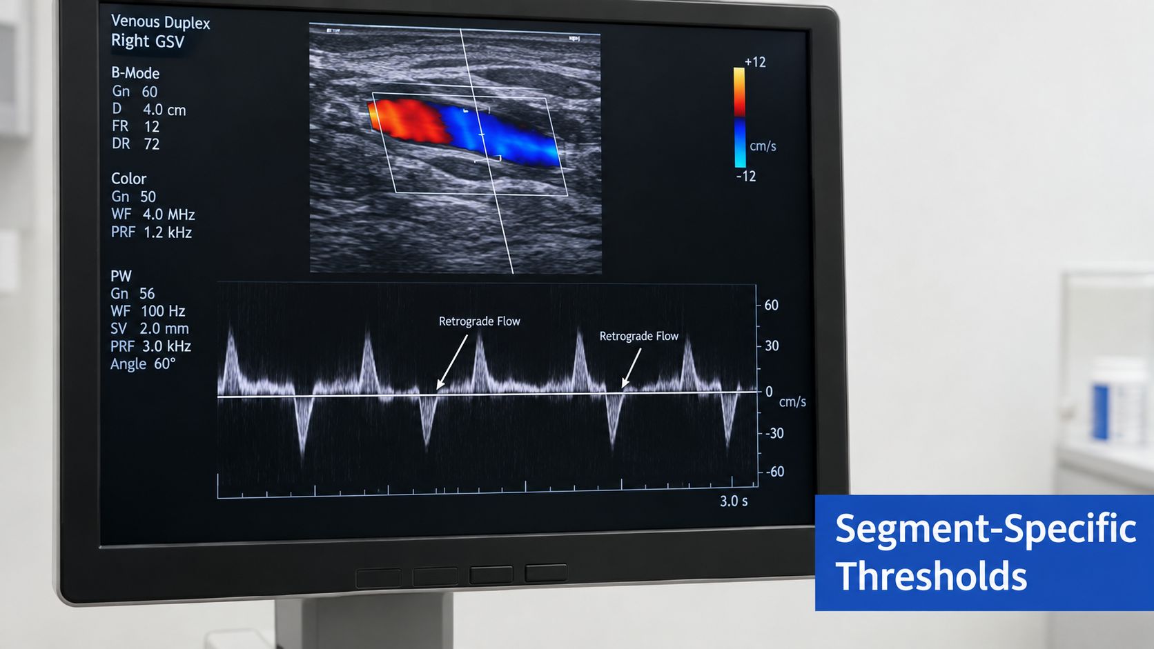
The number is only part of the answer
The specialist also considers:
- Location: Is reflux in the GSV, SSV, a deep vein, a perforator, or a tributary?
- Continuity: Does abnormal flow form a long pathway or remain confined to a short segment?
- Diameter: Is the vein enlarged at standardized measurement sites?
- Clinical effect: Do the findings fit the swelling, skin changes, pain, or wound location?
- Arterial safety: Could reduced arterial perfusion make compression or intervention unsafe?
A perforator larger than approximately 3.5 mm has been reported as associated with significant reflux, but diameter can't replace direct hemodynamic measurement (duplex assessment of venous reflux). The spectral trace remains important because it shows whether blood actually reverses and for how long.
Anatomy guides the treatment category
A continuous reflux pathway may support treatment directed at the incompetent axial source. Depending on the anatomy and the clinician's assessment, options can include endovenous laser ablation, radiofrequency ablation, adhesive closure, mechanochemical treatment, foam sclerotherapy, or ambulatory phlebectomy. A short segment or isolated tributary may instead lead to a more localized approach.
These names describe categories of procedures, not a recommendation for you. The map helps the specialist preserve healthy drainage while addressing the vein most responsible for symptoms.
A suspected venous ulcer requires an additional question: is arterial blood reaching the foot adequately? Arterial perfusion assessment, commonly with an ankle-brachial pressure index, may be needed before compression or invasive treatment. A technically precise venous map can't by itself prove that a wound is purely venous.
When a Normal Scan May Still Miss Venous Disease
You go to the emergency department with a swollen, painful leg. The ultrasound report comes back with reassuring words: “no evidence of DVT.” That result answers the urgent clot question. It may leave a second question open, whether faulty vein valves are allowing blood to fall backward and pool when you stand.
The gap usually begins with the order placed for the test. A scan requested for acute DVT often concentrates on whether the deep veins are open and compressible. Chronic venous disease requires a different setup, including reflux maneuvers, attention to superficial vein anatomy, and review of tributaries and perforators. As noted in teaching guidance on venous reflux assessment, unless the request specifically asks for superficial and deep valvular incompetence to be assessed, the examination may mainly address DVT.
Ask what the order is designed to find
For suspected chronic venous insufficiency, the request should clearly ask for a bilateral or clinically appropriate lower-extremity venous reflux study and name superficial and deep valvular incompetence. Depending on the symptoms, the clinician may also ask for mapping of the GSV, SSV, accessory veins, tributaries, and perforators.
A DVT-focused report can still be normal in someone with aching, heaviness, visible veins, swelling, or skin change from reflux. The earlier scan may have answered the question it was given, much like a chest X-ray can be useful while still leaving heart rhythm unexplored.
Why both legs can matter
Symptoms often draw attention to one leg, but a side-by-side study can add context. The other leg may show your usual anatomy, uncover disease on both sides, or help explain an asymmetric common femoral waveform when proximal obstruction is being considered.
If symptoms continue after a clot study, ask a concrete question: “Did this exam include a reflux evaluation of the superficial and deep systems, or do I need a dedicated venous reflux map?” Bring the original report. The named segments, patient position, and wording of the study often show whether another ultrasound would add useful information.
Mapping for Wounds, Diabetes, and Mixed Disease
A non-healing wound changes the purpose of vein mapping. The question is no longer only, “Which vein should be treated?” It becomes, “What is preventing this tissue from healing, and is compression safe?”
Consider an older patient with ankle swelling and an ulcer near the lower leg. Duplex mapping may show reflux in superficial and deep systems, an obstructed pathway, or a perforator connected to the wound region. International guidance identifies color-coded duplex assessment of both systems as the gold-standard method for evaluating these conditions before invasive treatment for venous leg ulcers (guideline evidence on venous leg ulcers).
A wound can have more than one cause
Venous disease may coexist with arterial insufficiency, diabetes, lymphedema, infection, edema, or pressure injury. Skin color and temperature, palpable pulses, ulcer location, drainage, pain pattern, and the pace of healing all add information that ultrasound can't replace.
If arterial compromise is possible, the clinician may order an ankle-brachial pressure index or toe-pressure testing before recommending compression. Compression is a standard part of venous-ulcer care, but it shouldn't be applied blindly when arterial flow may be reduced.
Diabetes adds another layer because reduced sensation can hide pressure or injury, while impaired circulation and infection can slow healing. A patient who wants an initial educational screen can get a diabetes risk score, but a score isn't a diagnosis and doesn't replace an examination, circulation testing, or wound evaluation.
The map supports a longer care plan
Suppose the scan identifies reflux, but the wound remains painful and the foot has weak pulses. Treating the superficial reflux alone may not solve the problem. The team may first clarify arterial status, control infection, protect the wound, and coordinate compression intensity with the circulation findings.
For patients with diabetes and lower-extremity wounds, diabetic leg ulcer care requires this broader view. The goal is not merely to label the ulcer. It is to identify the factors delaying closure, treat the circulation problem that can be corrected, and monitor the wound over time.
Mapping also creates a reference point for follow-up. If swelling changes, the ulcer reopens, or symptoms return, clinicians can compare the new examination with the earlier anatomy instead of starting from guesswork. Venous ulcers affect approximately 3% to 5% of adults aged 65 years and above, and approximately 50% to 76% reopen after healing, with recurrence risk increasing over subsequent years (medical literature on chronic venous disease and leg ulcers). That is why healing and prevention must be managed together.
Vein and Wound Institute evaluates venous disease and wound causes with clinical examination and duplex ultrasound, then coordinates minimally invasive vein treatment and advanced wound care when appropriate. If you have persistent symptoms, a non-healing wound, or a DVT-focused scan that didn't address reflux, visit Vein and Wound Institute to discuss whether a complete leg-vein map fits your care plan.