
The short answer is that ultrasound guided sclerotherapy is a needle injection into a target vein under live ultrasound, and the goal is to close that vein so blood reroutes through healthier veins. In the evidence base summarized by NICE, 84.4% was the median occlusion rate across five randomized trials involving 640 patients (NICE overview).
You're probably hearing those words in an exam room after a scan, while someone traces a vein on the screen and explains that the problem isn't just what's visible at the skin surface. This treatment is used when a vein is easy to find on ultrasound but hard to see or feel from the outside, which is why the phrase ultrasound guided matters so much. It's not a cosmetic quick fix in the simple sense, and it's not the same thing as treating every spider vein you can see.
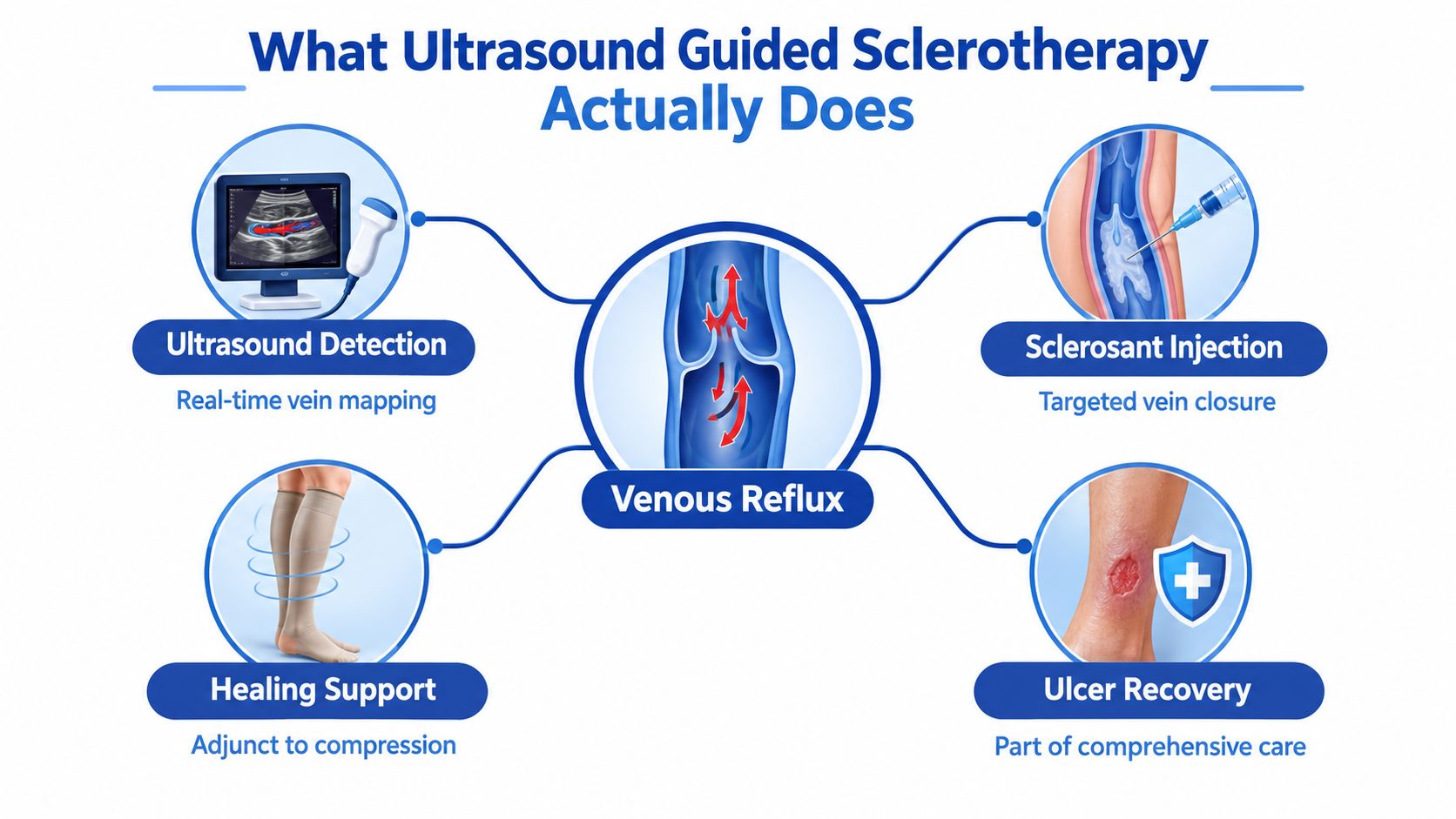
Table of Contents
- What Ultrasound Guided Sclerotherapy Actually Does
- How the Procedure Works Step by Step
- Realistic Outcomes and What the Numbers Mean
- How It Compares With Other Vein Treatments
- When Ultrasound Guided Sclerotherapy Fits a Vein Ulcer
- Recovery, Compression, and the Safety Profile
- When to See a Specialist and What to Ask
What Ultrasound Guided Sclerotherapy Actually Does
The moment usually feels very specific. A clinician has the ultrasound probe on your leg, the screen is showing a vessel you can't see from the outside, and now you're hearing about ultrasound guided sclerotherapy. What's happening is straightforward, even if the anatomy sounds intimidating, the team is using real-time ultrasound to find the right vein, place a small needle in the correct spot, and inject a sclerosant that irritates the vein lining until the vessel closes and is gradually reabsorbed.
Why ultrasound changes the procedure
That live imaging is what separates this from visual-only sclerotherapy. For veins near the skin surface, a clinician may treat by looking and feeling alone, but deeper or less obvious veins need a map. Ultrasound lets the clinician see where the vein runs, confirm that the needle is inside the vein, and watch the medication spread where it's supposed to go, rather than guessing.
Foam is often used when the vein is larger or deeper, because it has more surface contact with the vein wall than liquid alone. That matters when the target is a refluxing vein below the skin, not just a tiny surface spider vein. A practical rule is to think of it as image-guided vein closure, not just an injection.
Practical rule: if the vein can't be seen or reliably palpated, ultrasound is what turns treatment from approximate to targeted.
This is why the same procedure can mean different things for different patients. For one person, it's part of cosmetic varicose-vein care. For another, it's a way to treat a refluxing vein that's feeding recurrent symptoms or even contributing to a wound.
How the Procedure Works Step by Step
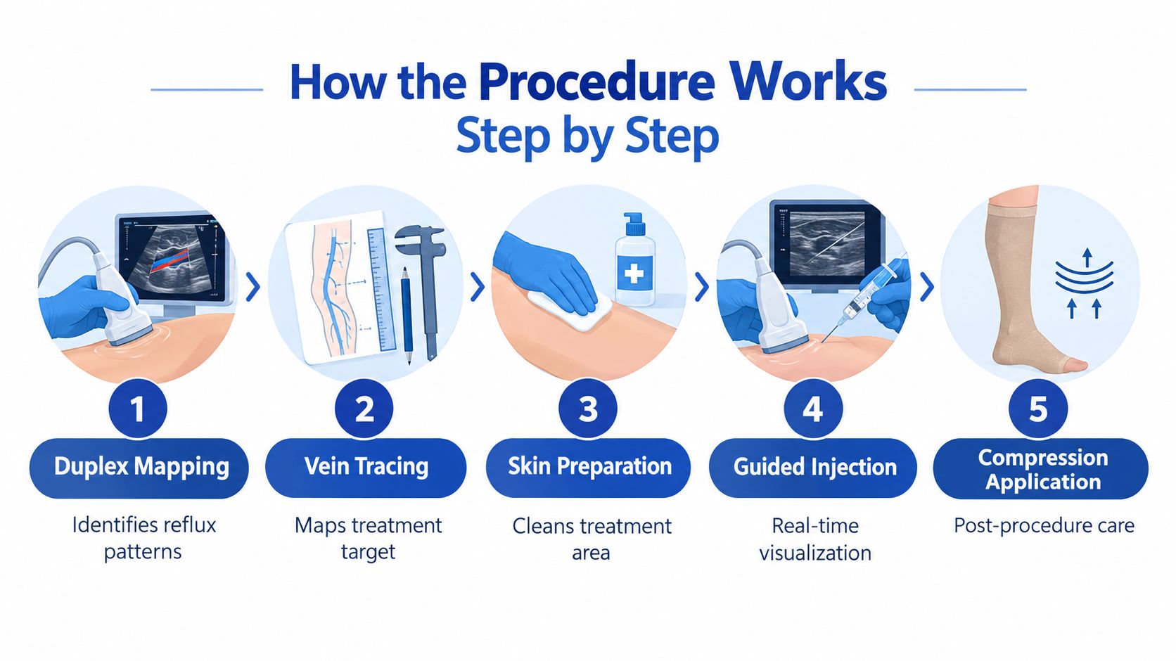
A patient may arrive expecting a simple injection, but the visit starts with a map. Duplex ultrasound shows the vein's route, confirms reflux, and helps the clinician avoid nearby arteries. It also shows whether the vein is a suitable target for this approach at all.
From mapping to injection
The sequence is straightforward once you break it down. The clinician identifies the target, places the needle under ultrasound guidance, injects the sclerosant in a controlled way, then scans again. That final look checks how the foam spread and whether any leakage occurred, so treatment stays in the intended vein.
Foam preparation has to be timed carefully. One technical reference recommends injecting within about 60 seconds because foam gradually destabilizes and shifts back toward a more liquid state (technical reference). That is why the room runs on a tight sequence, with scanning, mixing, and injection happening in close order.
What happens after the injection
The procedure does not end when the needle is removed. Compression is applied so the vein walls stay in contact while the sclerosant triggers inflammation and fibrosis. NICE describes the basic mechanism as endothelial injury followed by inflammatory fibrosis and venous blockage (NICE overview). In practical terms, the goal is to close a refluxing channel in a controlled way so the body stops using it.
This matters differently depending on the reason for treatment. In cosmetic varicose vein care, the aim is usually to reduce visible bulging and symptoms. In recurrent disease, the same method may target a residual refluxing segment that keeps feeding the problem. In venous leg ulcers, closing the wrong vein can interrupt the pressure that keeps the wound from healing. For a clear patient-focused walkthrough of the visit itself, this sclerotherapy procedure overview is a useful companion.
Realistic Outcomes and What the Numbers Mean
A patient may hear that a vein has been closed and assume the problem is finished. In real life, ultrasound guided sclerotherapy works more like one link in a longer vein-care plan. It can close a troublesome channel, reduce symptoms, and make future treatment easier, but the result depends on why the vein was treated in the first place.
For cosmetic varicose veins, success usually means the visible vein softens, shrinks, or disappears enough to improve appearance and comfort. For recurrent disease, the procedure may target a leftover refluxing segment that keeps feeding new varicosities. In venous leg ulcers, the goal is more functional, because closing the wrong pathway can leave pressure behind and slow healing.
The published numbers reflect that range of use. In the NICE summary of five randomized controlled trials involving 640 patients, the median venous-occlusion rate was 84.4%, with study results ranging from 67% to 94% across follow-up periods from 3 months to 10 years (NICE overview). That wide spread is a reminder that early closure and long-term durability are not the same thing. A vein can seal well at first and still reopen or be replaced by new reflux later.
A randomized study of 73 patients and 82 legs compared foam sclerotherapy plus saphenofemoral ligation with conventional surgery at 5 years. Above-knee obliteration was 58% after combined foam treatment versus 54% after surgery, and below-knee obliteration was 24% versus 39%, respectively. Neither difference was statistically significant, with p=0.19 above the knee and p=0.34 below the knee (NICE overview).
What those numbers mean in plain language
The treatment can work well, but no single percentage describes every patient. Anatomy matters, the treated vein matters, and the follow-up interval matters. A result that looks strong at the first check can look different years later if reflux returns or new veins become involved.
NICE also summarized a meta-analysis of two randomized trials including 340 patients, where foam was not significantly more effective than liquid sclerotherapy for vein occlusion. The relative risk was 1.5, with a 95% confidence interval of 0.6 to 3.6, and heterogeneity was very high at I²=95% (NICE overview).
Key takeaway: ultrasound guided sclerotherapy is best understood as a treatment that aims for closure, symptom relief, and planned follow-up, with recurrence kept in view rather than denied.
How It Compares With Other Vein Treatments
The best procedure is rarely the one with the loudest marketing. It's the one that matches the vein's shape, depth, reflux pattern, and your tolerance for downtime and repeat treatment. For tortuous, recurrent, or hard-to-access veins, ultrasound guided sclerotherapy often stays in the conversation because it can reach places that are awkward for other approaches.
Common minimally invasive vein treatments at a glance
| Treatment | Best fit | Typical downtime | Retreatment considerations |
|---|---|---|---|
| Ultrasound guided sclerotherapy | Nonvisible, tortuous, recurrent, or deeper veins | Usually limited, with compression often used | Repeat sessions may be needed if occlusion doesn't hold |
| Endovenous heat-based ablation | Straighter refluxing truncal veins | Often short, depending on anatomy and plan | May need adjunct treatment for branches or residual veins |
| Adhesive closure | Selected truncal reflux cases | Usually short | Retreatment depends on closure durability and residual disease |
| Injectable foam for visible tributaries | Surface branches and residual veins | Often limited | More than one session can be expected |
| Mechanochemical ablation | Great saphenous disease in selected patients | Procedure-specific | Comparative durability is still being studied |
| Ambulatory phlebectomy | Bulging visible branches | Brief recovery for many patients | Used when visible veins remain after deeper reflux is treated |
That comparison is intentionally practical. Patients usually want to know whether the vein is likely to be treated with local measures, whether compression will be expected, and whether the plan may involve more than one technique.
A registered randomized trial comparing ultrasound-guided foam sclerotherapy with mechanochemical ablation is designed to measure quality of life, ultrasound-confirmed occlusion, pain, and complications in 50 patients with great saphenous vein disease (trial record). That kind of design tells you the field is still refining the durability question rather than declaring a universal champion.
For a broader treatment-selection framework, this guide to choosing the right varicose-vein treatment is a helpful next read. If you're looking for a plain-language explanation of surface vein care, find spider vein removal guidance can also help you separate cosmetic concerns from deeper reflux questions.
When Ultrasound Guided Sclerotherapy Fits a Vein Ulcer
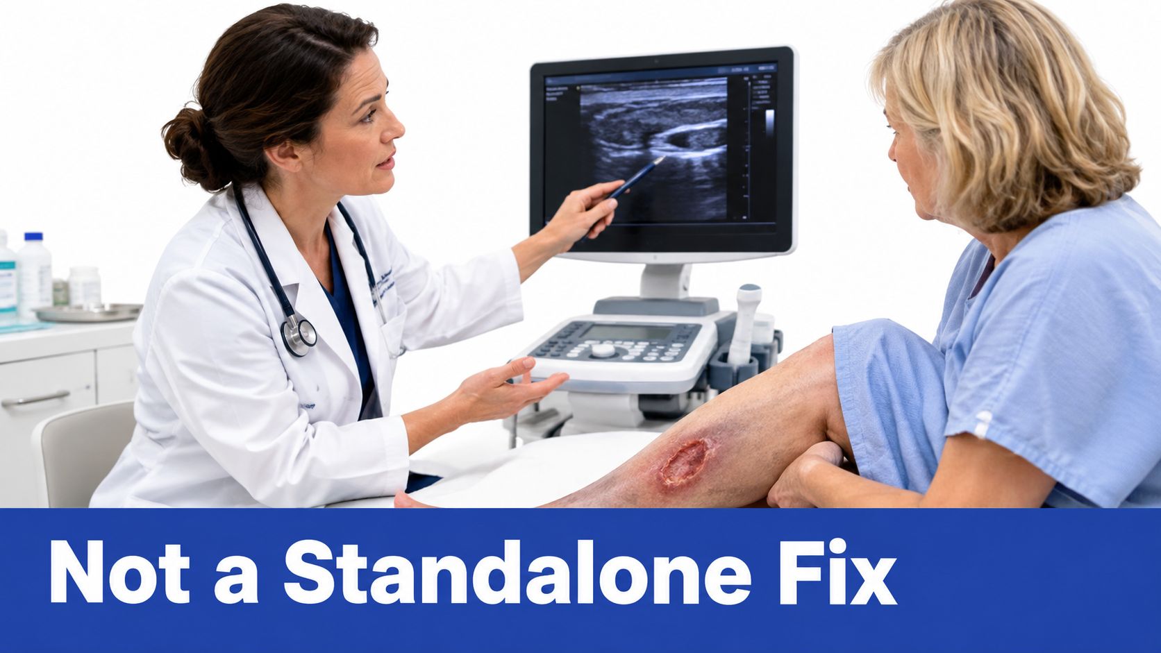
A venous leg ulcer changes the conversation completely. Sclerotherapy is never the whole wound treatment, because the wound still has to be managed with compression, local wound care, and an assessment of arterial supply. The procedure helps only when duplex ultrasound has shown that refluxing superficial or perforator veins are part of what's driving the ulcer.
Why the wound context matters
Patient-facing explanations often become too simplistic. If a wound is open, the question is not whether a vein looks closed on a scan, it's whether closing the reflux pathway changes the biology that keeps the ulcer from healing. That means treating the vein without treating edema, drainage, infection, pressure, or impaired arterial inflow misses the bigger clinical picture.
One review of advanced chronic venous insufficiency reported a 1-year pooled healing estimate of 99.9% for ultrasound-guided sclerotherapy of incompetent perforators, but it also described the underlying evidence as low-to-intermediate quality with meaningful risk of bias (advanced chronic venous insufficiency review). That combination matters. The number sounds impressive, but the review itself makes clear that the evidence is not clean enough to turn it into a blanket promise.
What to think about if the wound doesn't heal
A wound that stays open, or reopens after it seemed to close, may mean the vein plan wasn't complete. It can also point to persistent edema, infection, lymphedema, pressure, diabetes, or arterial disease. In a vein-and-wound practice, the value of ultrasound guided sclerotherapy is that it can remove one major driver of venous hypertension while the rest of the wound program keeps working.
For ulcers, the right question is not “Did the injection work?” It's “Did the overall reflux-and-wound strategy finally remove the barrier to healing?”
A doctor reviewing a wound alongside duplex imaging is looking for that larger answer, not a cosmetic result. That's why venous ulcer care and vein closure should be planned together, not treated as separate problems that happen to share a leg.
Recovery, Compression, and the Safety Profile
The first days after treatment are usually routine rather than dramatic. Compression starts right away, and the exact duration depends on the protocol and the treated anatomy. Walking is usually encouraged, because calf movement helps venous return without the strain of hard exercise. For a plain-language overview of home care, this recovery guide is useful.
What to expect at home
Bruising, tightness, and a lumpy feel along the treated vein are common and usually fade gradually. Cosmetic change often trails behind the procedure, because the vein has to close, break down, and then be absorbed. A patient may feel the vein is still there for a while, even when the treatment is doing its job.
The safety profile is reassuring, but not zero-risk. In the 2007 systematic review, the median frequency of serious adverse events, including deep-vein thrombosis and pulmonary embolism, was below 1% (BJS review). The same review reported a median recurrence or new-vein rate of 8.1%, which is one reason follow-up remains part of vein care rather than an afterthought.
For some patients, that follow-up is about appearance. For others, it is about recurrent disease or keeping an ulcer plan on track. The procedure plays a different role in each setting, but the need to watch the leg over time is the same.
When to call the clinic
- Increasing leg swelling: especially if it is new or clearly worse than expected.
- Calf pain: particularly if it is persistent or paired with swelling.
- Chest discomfort: this needs prompt evaluation.
- Skin color changes at the injection site: worsening discoloration or concerning redness should be checked.
- Visual disturbances: do not ignore these, even if they seem temporary.
When to See a Specialist and What to Ask
If your legs ache, swell, or throb enough to change how you get through the day, it is time for a specialist visit. The same applies when swelling does not settle overnight, the skin near the ankles darkens, a venous leg ulcer or other wound is slow to heal, varicose veins come back after prior treatment, or venous disease runs in the family. Those are the moments when a duplex-informed consultation matters more than another guess based on how the leg looks.
Ultrasound guided sclerotherapy is one part of a longer vein-care plan. For cosmetic varicose veins, it may close a visible problem that bothers appearance and symptoms. For recurrent disease, it can target a refluxing segment that was missed, reopened, or developed later. For venous ulcers, it may support wound healing by reducing the pressure from abnormal flow, but the ulcer plan still needs follow-up, compression, and reassessment over time.
Questions that help you get a useful plan
- Which veins are being treated, and why? Ask for the reflux source, not just the visible branch.
- What follow-up ultrasound is planned? The answer should include surveillance, not only the procedure day.
- What compression will I need? Ask how long, how often, and why your plan differs from someone else's.
- What happens if the vein does not stay closed? Retreatment is part of real-world vein care, so the fallback plan should be clear.
- How does this fit with diabetes, arterial disease, or an active wound? Those factors change timing and priorities.
If the clinic uses a patient intake coordinator, that person should help gather records, symptoms, prior procedure history, and wound details before the visit. That lets the specialist spend more time on the decision itself and less time rebuilding the story from scratch.
A good vein consultation should feel like a plan that continues after the first visit. Ultrasound guided sclerotherapy fits that kind of care best when recurrence prevention and wound healing both matter.
Vein and Wound Institute treats venous disease and chronic wounds together, which matters when ultrasound guided sclerotherapy is being considered as part of a broader plan. If you're trying to understand whether a refluxing vein, a recurrent varicose vein, or a venous ulcer should be addressed first, visit Vein and Wound Institute to learn more about coordinated vein and wound care.