
Red or purple discoloration on the legs isn't automatically bruising, a harmless skin mark, or cellulitis. It can reflect pooled venous blood, chronic inflammation, infection, clot-related obstruction, or reduced arterial flow. The color matters, but the surrounding clues matter more: when it started, whether one or both legs are affected, whether the skin is warm, whether swelling or pain is present, whether pulses are reduced, and whether an ulcer or other wound has appeared.
A sudden change in one leg with swelling, warmth, or pain needs prompt medical assessment. Fever with rapidly spreading redness, shortness of breath with leg symptoms, severe pain, a cold foot, or darkening tissue raises the urgency further. Clinicians may use a physical examination, duplex ultrasound, and an ankle-brachial index, or ABI, to distinguish venous disease from arterial insufficiency, thrombosis, and infection. Appearance alone can't establish the diagnosis.
The following seven causes are separated by the clues that help point toward the right next step. Don't start high-pressure compression on unexplained red purple skin on legs until arterial circulation has been assessed.
Table of Contents
- 1. Chronic Venous Insufficiency with Venous Hypertension
- 2. Varicose Veins with Secondary Skin Changes
- 3. Lipodermatosclerosis as Severe Venous Skin Change
- 4. Hemosiderin Staining and Venous Dermatitis
- 5. Deep Vein Thrombosis and Post-Thrombotic Syndrome
- 6. Arterial Insufficiency and Peripheral Artery Disease
- 7. Cellulitis and Skin Infection
- Red–Purple Leg Skin: 7-Condition Comparison
- Match the Color Pattern to the Right Next Step
1. Chronic Venous Insufficiency with Venous Hypertension
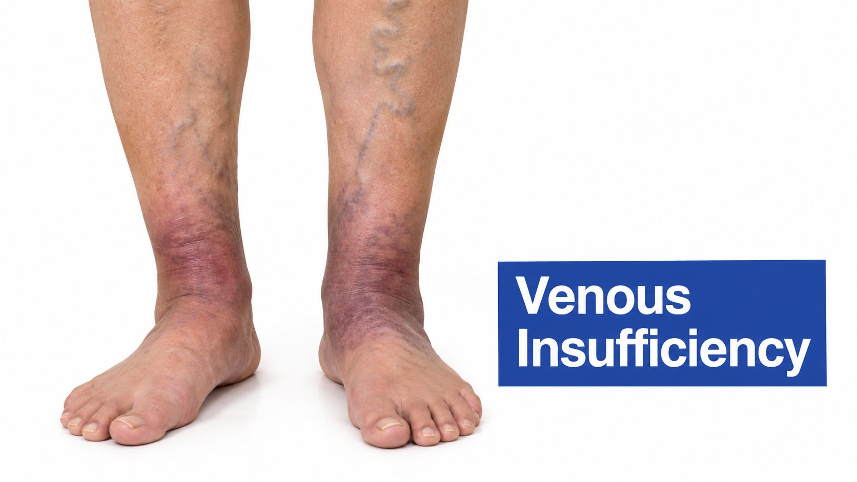
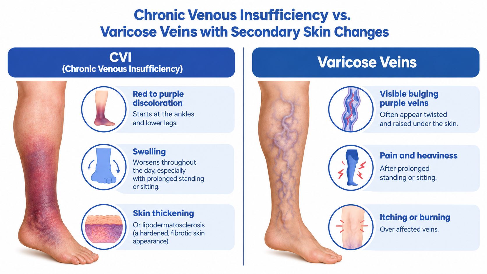
Chronic venous insufficiency, or CVI, occurs when leg veins cannot move blood back to the heart efficiently. Blood pools in the lower limbs, venous pressure rises, and skin around the ankles or lower legs may become red, purple, brown, thickened, or irritated. The color often becomes more visible after standing and may occur with leg heaviness, swelling, aching, itching, or visible veins.
CVI affects more than 25 million adults in the United States. It can progress from reticular or varicose veins to edema, pain, hyperpigmentation, hardened skin, and ulceration, as described in the National Center for Biotechnology Information clinical overview of chronic venous disease. An ulcer indicates tissue damage and requires more than cosmetic treatment.
What separates CVI from other causes
CVI usually develops gradually and often affects both legs, although one side may be worse. Swelling that builds during the day and eases with elevation supports a venous cause. Duplex ultrasound can assess reflux or obstruction, while pulse examination and an ankle-brachial index help identify arterial disease. A sudden, painful, warm, one-sided change needs assessment for DVT rather than routine CVI care. Fever and rapidly spreading redness point toward cellulitis.
Practical rule: Compression can reduce venous swelling and support ulcer healing, but arterial perfusion must be assessed first. Weak pulses, a cold foot, rest pain, or a nonhealing wound make unsupervised compression unsafe.
For venous ulcers, compression should follow arterial assessment, accurate measurement, pressure selection, fitting, and monitoring. A 2023 meta-review of compression for venous leg ulcers found healing in 61% of participants receiving compression compared with 39% without compression. Pain, numbness, color change, or pressure injury calls for prompt review.
Treatment may also address reflux, edema, and wound care rather than skin color alone. Topical support can help irritated skin, but it does not correct venous hypertension. See MEDISTIK product pairing explained for product-use guidance. Patients can also review how to spot venous insufficiency and when to visit a vein treatment center.
2. Varicose Veins with Secondary Skin Changes
Varicose veins are enlarged, twisted superficial veins that become visible when valves allow blood to move backward and pool. The veins themselves may look blue or purple, while nearby skin develops red-purple or brown discoloration as venous pressure affects small vessels and surrounding tissue.
The visual finding is easy to dismiss as cosmetic, especially when there isn't much pain. That can be a mistake. A patient may report aching, heaviness, itching, ankle swelling, or discomfort after prolonged standing even when the main concern is the appearance of the veins.
Look beyond the visible vein
A useful clinical question is whether the discoloration sits around a cluster of varicosities or extends toward the ankle, where chronic venous pressure can produce dermatitis, staining, or ulcer risk. A duplex ultrasound can identify reflux in a larger superficial vein and show whether the visible branches are part of a broader circulation problem.
Treatment should follow the anatomy. If a major refluxing vein is driving pressure, closing that source with an endovenous technique may be more logical than treating every small visible branch first. Residual veins can then be considered for sclerotherapy or ambulatory phlebectomy, depending on their size, location, and symptoms. Cosmetic treatment without addressing significant reflux may leave the underlying pressure unchanged.
The American Academy of Family Physicians review of compression and venous ulcer recurrence describes a trade-off that applies broadly to venous care: stronger compression can provide more protection against recurrence, but patients may find it harder to wear consistently. The best plan is one a patient can use correctly and sustain.
Visible veins are a clue, not a complete diagnosis. The surrounding swelling, skin texture, ulcer history, and arterial examination determine the safe treatment direction.
A patient with painful or enlarging varicosities should receive an assessment rather than relying only on topical products or over-the-counter hosiery. The available treatment options for varicose veins range from conservative management to minimally invasive procedures, selected after the cause of the reflux is established.
3. Lipodermatosclerosis as Severe Venous Skin Change
Lipodermatosclerosis is an advanced tissue response to sustained venous hypertension. Inflammation and fibrosis make the skin and underlying fat firm, tight, and less flexible. The typical pattern is a red-purple, painful, hardened band around the lower calf, often near the inner ankle, with narrowing above the ankle that can create an inverted-champagne-bottle contour.
The texture helps separate it from simple discoloration. The area may feel woody or tender, and minor trauma can cause disproportionate skin breakdown. Sudden warmth, rapidly increasing pain, marked swelling, or fever points toward infection or an acute venous problem instead of stable lipodermatosclerosis and requires prompt assessment.
The warning signs of progression
Suspected lipodermatosclerosis warrants evaluation for venous reflux, edema, and an ulcer that may be developing beneath the altered skin. Watch for new warmth, weeping, increasing tenderness, a break in the skin, or drainage. These findings may signal ulceration or infection and should not be treated as a cosmetic issue.
Duplex ultrasound can assess reflux and help determine whether vein-directed treatment has a role. Before high-pressure compression is prescribed, check arterial supply with pulse examination and an ankle-brachial index or another appropriate arterial study. Compression can support venous return, but significant arterial insufficiency changes its safety and intensity.
Chronic venous disease may progress from discoloration and tissue hardening to ulceration. Venous leg ulcers are the most common type of chronic lower-extremity wound, and approximately 1% to 3% of older adults in the United States and Europe are affected, according to the clinical review of venous disease and ulceration. A wound near a discolored ankle therefore deserves circulation testing rather than routine self-treatment.
Management aims to reduce venous pressure, protect fragile skin, support healing, and limit recurrence. This may involve appropriately selected compression, wound care, edema control, and treatment of confirmed reflux. Follow-up should track swelling, pain, skin integrity, and ulcer development. Treating an open wound while leaving major venous pressure unchecked may allow the tissue damage to continue.
4. Hemosiderin Staining and Venous Dermatitis
Hemosiderin staining is a chronic color change caused by red blood cells leaking from pressurized, fragile veins. As those cells break down, iron-containing pigment remains in the skin, creating brown, rust-colored, red-purple, or mottled patches. The pattern commonly appears around the ankles and lower legs, where venous pressure and swelling tend to be greater.
Venous dermatitis may develop in the same area. It causes itching, scaling, cracking, redness, or inflammation. Scratching can create breaks in the skin and increase irritation or infection risk, especially when edema has weakened the skin barrier.
Separate pigment from active inflammation
Staining usually represents residual pigment. Dermatitis is active inflammation, and one patient may have both. Examine the skin for warmth, tenderness, weeping, or a new break. Ask whether the color changes with leg position, and assess pulses when arterial disease is possible. These findings help distinguish venous disease from arterial insufficiency, bruising, pigmented purpura, and infection.
Orange-red-brown patches with pinpoint spots can also occur in pigmented purpuric conditions. They may resemble venous hypertension, but the causes and treatment differ. The vascular guidance on chronic venous disease and compression explains why discoloration can have several mechanisms. Do not begin firm compression until arterial supply has been assessed with pulse examination and an ankle-brachial index or another suitable arterial test.
For additional context on differentiating pigmented purpura from venous dermatitis, see Outrank's dermatologist playbook.
Treat the circulation problem and the irritated skin separately. Moisturizer or a prescribed topical anti-inflammatory may calm dermatitis, but it will not correct venous reflux.
If reflux is confirmed and arterial flow is adequate, clinicians may combine selected compression with vein treatment and skin care. Color improvement is often slow, and established pigment may remain. Avoid harsh scrubbing, unprescribed antibiotic ointments, and scratching. A wound, drainage, or rapidly changing appearance requires clinical examination rather than cosmetic treatment.
5. Deep Vein Thrombosis and Post-Thrombotic Syndrome
Deep vein thrombosis, or DVT, occurs when a clot blocks flow in a deep leg vein. The acute pattern is usually more concerning than longstanding staining: one-sided swelling, pain, warmth, and a new change in color should prompt urgent assessment. A person may not have every symptom, so the absence of dramatic pain doesn't safely rule out a clot.
Duplex ultrasound is commonly used when DVT is suspected because it can evaluate deep venous flow and obstruction. The immediate priority is confirming the diagnosis and protecting the patient from complications, including clot migration to the lungs. Shortness of breath, chest pain, coughing blood, fainting, or sudden unexplained breathlessness with leg symptoms requires emergency care.
Chronic changes after a clot
A previous DVT can damage valves and leave residual obstruction. Over time, the affected leg may develop persistent swelling, heaviness, red-purple or brown discoloration, dermatitis, and ulceration. This chronic pattern is different from an acute clot, but it still requires a circulation plan.
Clinicians may use compression after assessing the patient and may address confirmed reflux or obstruction once the acute phase has been managed. A procedure aimed at superficial veins shouldn't be chosen just because the leg looks discolored. The medical history, current anticoagulation plan, repeat symptoms, and ultrasound findings all matter.
A patient who notices a sudden difference between legs should record when it began and whether activity, elevation, or rest changes the symptoms. Don't massage a potentially clotted leg or delay evaluation while trying home remedies. Likewise, don't assume that every long-term purple mark represents a new DVT. The time course and examination distinguish an acute event from post-thrombotic skin and circulation changes.
6. Arterial Insufficiency and Peripheral Artery Disease
Arterial insufficiency reduces blood delivery to the leg and foot. The skin may look pale, shiny, or cool, but advanced or positional disease can also produce red-purple discoloration. The most important clues are often not the color. They include weak or absent pulses, slow-healing sores, reduced hair growth, coldness, exertional leg discomfort, or pain at rest.
Peripheral artery disease can be clinically important even without classic exertional pain. The Centers for Disease Control and Prevention overview of peripheral artery disease reports that up to 4 in 10 people with PAD have no leg pain, so a comfortable walk doesn't exclude reduced arterial flow. PAD may also cause foot pulses to be decreased or absent and wounds on the legs or feet that fail to heal.
Why compression can be dangerous
Venous disease and arterial insufficiency require different treatment decisions. Compression can support venous return, but high pressure may worsen tissue ischemia when arterial inflow is inadequate. That's why ABI testing and pulse assessment should precede high-pressure compression when arterial disease is possible.
A patient with a cold foot, absent pulses, rest pain, tissue loss, or a non-healing foot ulcer needs arterial assessment and, when indicated, vascular referral. Duplex imaging may help define blood flow, while additional testing depends on the clinical findings. Do not treat purple skin as a venous problem because it appears near the ankle.
The causes of arterial leg ulcers illustrate why wound location, pain, temperature, and pulses matter. A wound-care plan that skips circulation testing can miss the reason tissue isn't healing.
7. Cellulitis and Skin Infection
Cellulitis is an acute bacterial infection involving the deeper skin and subcutaneous tissues. It typically produces redness, warmth, swelling, and tenderness, often with a clear area of progression. Fever or feeling unwell increases concern, but infection can still require assessment when systemic symptoms are absent.
The key distinction is speed and behavior. Cellulitis tends to develop or spread over a short period, while venous dermatitis is more often gradual, itchy, scaly, and recurrent around swollen ankles. Red-purple discoloration by itself can't identify cellulitis. Stasis dermatitis, erysipelas, superficial or deep venous thrombosis, gout, contact dermatitis, and other inflammatory conditions can look similar, as outlined in the evidence-based dermatology guidance on the red leg differential.
When infection needs prompt care
Rapidly spreading warmth, marked tenderness, fever, pus, skin breakdown, or a sudden deterioration needs prompt medical evaluation. Antibiotics may be required, and delaying care while trying compression, steroid cream, or cosmetic products isn't appropriate when infection is suspected.
Chronic venous disease can increase infection risk because swelling and dermatitis weaken the skin barrier. Small cracks between the toes, fungal skin disease, scratching, and ulcers can provide entry points for bacteria. After an acute infection is treated, clinicians should look for the factors that allowed it to develop, including edema, venous insufficiency, lymphedema, dermatitis, and open wounds.
Don't label every red leg as cellulitis, but don't dismiss rapidly changing warmth and pain as “just circulation” either.
Compression may have a role in managing underlying venous edema, but it should be introduced according to the examination and arterial status. A patient with a suspected infection needs medical treatment first, followed by a plan to protect the skin and address the circulation problem that may be contributing to recurrence.
Red–Purple Leg Skin: 7-Condition Comparison
| Condition | Implementation complexity | Resource requirements | Expected outcomes | Ideal use cases | Key advantages |
|---|---|---|---|---|---|
| Chronic Venous Insufficiency (CVI) with Venous Hypertension | Moderate, requires duplex mapping, long-term follow-up and possible staged ablation | Duplex ultrasound, compression therapy, EVLA/RFA/VenaSeal/foam sclerotherapy, outpatient clinic resources | Symptom relief and reduced ulcer risk; chronic management with possible recurrence | Patients with ankle/lower-leg discoloration, edema, varicose veins and documented reflux | Multiple minimally invasive outpatient options; immediate relief from compression; prevents ulcer progression |
| Varicose Veins with Secondary Skin Changes | Low–moderate, outpatient procedures often suffice; duplex assessment recommended | Duplex ultrasound, ablation or sclerotherapy devices, phlebectomy instruments, compression | Rapid symptomatic and cosmetic improvement; prevents progression if reflux treated | Bulging superficial veins with heaviness/cramping or cosmetic concern | Good cosmetic results; same-day procedures; quick symptom relief |
| Lipodermatosclerosis (LDS), Severe Venous Skin Changes | High, urgent multidisciplinary evaluation, aggressive compression and wound prevention | Urgent duplex, ABI, high-compression therapy, wound care resources, possible ablation | Slows progression and lowers ulcer risk if treated early; some changes may be irreversible | Indurated inverted‑champagne leg, firm medial ankle band, high risk for ulcers | Recognition triggers urgent, disease‑modifying care; integrated vein and wound management reduces complications |
| Hemosiderin Staining and Venous Dermatitis | Low–moderate, clinical diagnosis with duplex to confirm underlying reflux; prolonged therapy | Duplex ultrasound, compression stockings, topical corticosteroids/emollients, skin care | Partial to full cosmetic improvement (best when early); dermatitis control over months | Brown/red pigmentation with itching/scaling on medial ankle and history of venous insufficiency | Often reversible when treated early; combination of topical therapy + compression effective |
| Deep Vein Thrombosis (DVT) with Post‑Thrombotic Syndrome (PTS) | High, acute emergency management followed by complex chronic care | Duplex ultrasound, anticoagulation (DOAC/heparin), compression, possible thrombolysis or specialty interventions | Acute clot control; variable long‑term PTS outcomes; often lifelong compression; recurrence risk exists | Acute unilateral painful swollen leg or patients with prior DVT developing chronic venous disease | Anticoagulation prevents extension/PE; duplex reliably diagnoses; selected interventions can improve chronic symptoms |
| Arterial Insufficiency (Peripheral Arterial Disease) with Skin Changes | Moderate–high, requires arterial testing and specialist referral; mismanagement risk if confused with venous disease | ABI, arterial duplex/angiography, endovascular/surgical teams, medical risk‑factor management | Improved perfusion after revascularization; poor healing if untreated; compression contraindicated in moderate‑severe disease | Claudication, rest pain, absent pulses, non‑healing foot/toe wounds or atherosclerotic risk factors | Clear diagnostic tests (ABI); revascularization can prevent limb loss; medical and exercise therapies improve outcomes |
| Cellulitis and Skin Infection (Acute vs Chronic Dermatitis) | Variable, acute cellulitis is high urgency; chronic dermatitis is low–moderate complexity | Clinical assessment, antibiotics (oral/IV), blood cultures/imaging if severe, wound care; duplex to assess underlying venous disease | Acute cellulitis usually resolves rapidly with antibiotics; recurrent cases need venous evaluation and prophylaxis | Acute red, warm, tender swollen leg with systemic signs; recurrent infections in patients with venous disease | Prompt antibiotics give fast resolution; treating underlying venous disease reduces recurrence; clear clinical pathways for acute care |
Match the Color Pattern to the Right Next Step
Red purple skin on legs needs triage by pattern, timing, and associated symptoms, not by color alone. The safest next step changes depending on whether the problem is sudden or longstanding, one-sided or bilateral, warm or cool, painful or itchy, and associated with swelling, fever, a wound, or breathing symptoms.
Use these practical distinctions:
- Sudden one-sided swelling, warmth, or pain: Seek prompt medical evaluation for possible DVT or infection. Shortness of breath, chest pain, fainting, or sudden breathlessness raises the urgency to emergency care.
- Fever with rapidly spreading warmth: Arrange prompt infection care. Don't wait for a cosmetic explanation or attempt to manage a suspected cellulitis episode with compression alone.
- Cold foot, absent or weak pulses, rest pain, or tissue loss: Request urgent arterial assessment. These findings can indicate inadequate blood flow, even when the leg isn't severely painful during walking.
- Gradual ankle discoloration with swelling, heaviness, varicosities, itching, or ulcers: Arrange a venous evaluation. Chronic venous disease can progress from visible skin changes to hardened tissue and ulceration.
- Persistent pinpoint spots or net-like purple patterns: Ask for a broader differential diagnosis. Non-blanching purpura, livedo reticularis, bruising, and small-vessel disorders don't have the same treatment as venous hypertension.
Clinicians may examine pulses, edema, temperature, skin integrity, and positional changes before ordering duplex ultrasound. Duplex imaging can assess venous reflux, obstruction, and suspected thrombosis. ABI testing helps determine whether arterial flow is adequate before high-pressure compression or a venous ulcer plan is started.
Compression is useful for appropriately selected venous disease, but it isn't a universal answer for discoloration. The intervention must match the mechanism. A venous ulcer may benefit from structured compression after arterial disease is excluded, while an ischemic foot needs arterial evaluation and a different treatment direction.
Before your appointment, photograph the affected area in consistent lighting and note when the change began, whether it is spreading, and whether swelling or pain changes with standing or elevation. Don't scratch fragile skin, apply strong topical products to an open wound, massage a suddenly swollen leg, or self-treat an unexplained ulcer. Vein and Wound Institute can be relevant when suspected venous disease and active wounds need coordinated diagnostic, vein, and wound care, including examination and duplex ultrasound.
Vein and Wound Institute evaluates venous disorders, leg swelling, varicose veins, venous leg ulcers, arterial leg ulcers, diabetic wounds, and other non-healing wounds through coordinated vein and wound services. If your discoloration is persistent, worsening, or associated with swelling or skin breakdown, schedule an assessment by visiting Vein and Wound Institute.