The popular advice says to treat varicose veins as a cosmetic nuisance and deep vein thrombosis as an emergency that belongs in a completely different box. That framing is too simple for bedside care. In practice, the question is not whether the two conditions are “related” in theory, but whether visible varicosities are signaling a higher clot-risk profile that deserves imaging now rather than later.
| Feature | Varicose Veins | Deep Vein Thrombosis |
|---|---|---|
| Primary problem | Chronic superficial venous reflux | Acute clot in the deep venous system |
| Typical pace | Develops gradually | Often appears suddenly or worsens over hours to days |
| Main bedside concern | Symptoms, skin change, chronic venous disease | Clot propagation and possible pulmonary embolism |
| First-line imaging | Duplex ultrasound for reflux mapping | Duplex ultrasound for non-compressible thrombus |
| Urgency | Often elective unless symptoms change | Often urgent when symptoms are unilateral or acute |
Practical rule: a visible vein problem is not automatically “just cosmetic,” and a painful swollen leg is not automatically “just varicose veins.” The bedside task is to decide whether the pattern fits chronic reflux, an acute clot, or both.
Table of Contents
- Why Varicose Veins and DVT Are Not the Same Conversation
- What Each Condition Actually Means
- Causes and Risk Factors Compared
- Symptoms That Help You Tell Them Apart
- How Much DVT Risk Varicose Veins Really Signal
- Diagnosis and When to Get Imaging
- Treatment Paths for Each Condition
- When to Seek Urgent Care Versus a Vein Evaluation
Why Varicose Veins and DVT Are Not the Same Conversation
The easiest way to miss the point is to compare these conditions as if they sit at opposite ends of a simple severity scale. They don't. Varicose veins are common chronic venous disease, affecting roughly 23% of U.S. adults, and estimates rise when spider telangiectasias and reticular veins are included (AHA review). DVT is much less common, but it carries far higher acuity because the clot can extend or embolize.
The bedside question is risk, not labels
A patient can have obvious ropey veins and still develop a deep clot. That's why the practical question is not “Which one is this?” in isolation. It's “Does this patient's pattern suggest simple chronic reflux, or do the symptoms and context justify urgent evaluation for thrombosis?”
A leg with bulging surface veins can still hide a deep clot. If the symptoms are unilateral, new, painful, or rapidly changing, treat the presentation as a thrombotic problem until proven otherwise.
Many patient-facing explanations fall short. They describe varicose veins as benign and DVT as separate, then stop there. At the bedside, the distinction matters less than the triage decision. A chronic bulge that has been stable for years belongs on a scheduled vein workup. A leg that changes overnight does not.
What Each Condition Actually Means
Varicose veins are dilated, tortuous superficial veins caused by chronic valve failure and reversed flow. They usually involve the saphenous system or connected tributaries, so the problem sits in the superficial venous network rather than the deep system. DVT, by contrast, is an acute thrombus inside a deep vein, often in the femoral, popliteal, or iliac segments, with possible embolic travel to the lungs.
Anatomical hinge: superficial reflux creates visible surface veins, while deep venous clot creates an embolic risk problem. That single difference drives urgency, testing, and treatment.
Duplex ultrasound is the key study for both conditions because it can map reflux anatomy in varicose veins and check for a non-compressible thrombus in suspected DVT (PMC review). It's the same imaging tool, but the question changes. For varicose veins, the scan asks where the reflux is coming from. For DVT, it asks whether the vein compresses normally.
If you want a plain-language patient reference on clot symptoms and escalation, the internal DVT resource is a useful companion to the clinical distinction.
Why the anatomy matters
A superficial vein can be unsightly, uncomfortable, and progressive without being an emergency. A deep clot may be less visible but much more dangerous. That's why the same leg can contain both chronic reflux and a new thrombotic event, especially when symptoms abruptly change.
Causes and Risk Factors Compared
Varicose veins and DVT overlap in some risk factors, but they arise from different mechanisms. Varicose veins develop from chronic reflux, often tied to hereditary valve incompetence, prolonged standing, pregnancy, obesity, female sex, and age-related weakening of the vein wall. DVT develops from Virchow's triad, venous stasis, endothelial injury, and hypercoagulability, which is why immobility, recent surgery, cancer, hormone therapy, thrombophilia, and active inflammation matter so much.
| Risk Factor | Varicose Veins | DVT |
|---|---|---|
| Prolonged standing | Common contributor | Can contribute through stasis, but not a primary driver alone |
| Pregnancy | Common contributor | Can increase thrombosis risk through hypercoagulability and stasis |
| Obesity | Common contributor | Common contributor |
| Immobility | Can worsen venous symptoms | Major driver through stasis |
| Recent surgery | Not a classic cause | Major driver through endothelial injury and stasis |
| Cancer | Not a classic cause | Major driver through hypercoagulability |
| Inherited thrombophilia | Not a typical cause | Important risk factor |
| Age | Increases venous disease burden | Increases thrombotic risk |
Shared inputs deserve respect
Age, obesity, and immobility appear on both lists, and that overlap is the whole point. They don't mean the same thing in both conditions, but they do create a shared vulnerability in the venous system. That's why visible varicosities should never be dismissed as a purely cosmetic finding when the history includes new swelling, pain, or recent immobilization.
Clinical takeaway: the same patient can have chronic reflux and then develop a clot on top of it. The right question is which risk factor is active right now, and whether the current symptoms fit a clotting event.
Some factors are modifiable, some aren't. Weight control, movement, and avoiding prolonged immobility can help both the chronic reflux picture and the clot-risk picture. Family history, age, and inherited thrombophilia can't be changed, but they should lower the threshold for imaging when symptoms shift.
Symptoms That Help You Tell Them Apart
The symptom pattern usually gives the first clue. Varicose veins commonly cause heaviness, aching, visible bulging, evening ankle swelling that eases with elevation, and itching or burning over a ropey vein. If you need a fuller symptom pattern, see our guide to varicose vein symptoms. DVT more often causes one-sided calf or thigh pain, swelling in one leg, warmth, redness, and tenderness that feels out of proportion to what the skin looks like.
The four bedside axes that matter
Laterality matters. Varicose vein symptoms are often bilateral or show the same chronic asymmetry patients have had for years. DVT is more likely to appear on one side, especially when the leg is larger, warmer, or more discolored than the other.
Speed matters too. Chronic reflux builds gradually. DVT often develops over hours to days, or after a period of stillness. Tenderness quality matters as well, because clot pain is often deeper and less tied to a visible ropey vein. Color change matters, because new redness or dusky discoloration in one leg deserves more than reassurance.
For a practical patient-facing checklist, the warning signs of DVT guide is useful when symptoms change suddenly.
| Clinical Cue | Varicose Veins | DVT |
|---|---|---|
| Laterality | Often bilateral or long-standing asymmetry | Commonly unilateral |
| Pain quality | Dull ache, heaviness, burning | Deeper, sharper, or disproportionate |
| Swelling pattern | Mild, often worse by evening | One-leg swelling, often abrupt |
| Skin color | Chronic discoloration near ankles | New redness or dusky change |
| Onset | Gradual | Sudden or progressive over hours to days |
Subtle presentations happen. Roughly half of DVT cases can look subtle or atypical, so a normal-looking leg does not rule it out. The combination that should raise concern is unilateral swelling, pain, warmth, and discoloration, especially when the symptoms are new.
How Much DVT Risk Varicose Veins Really Signal
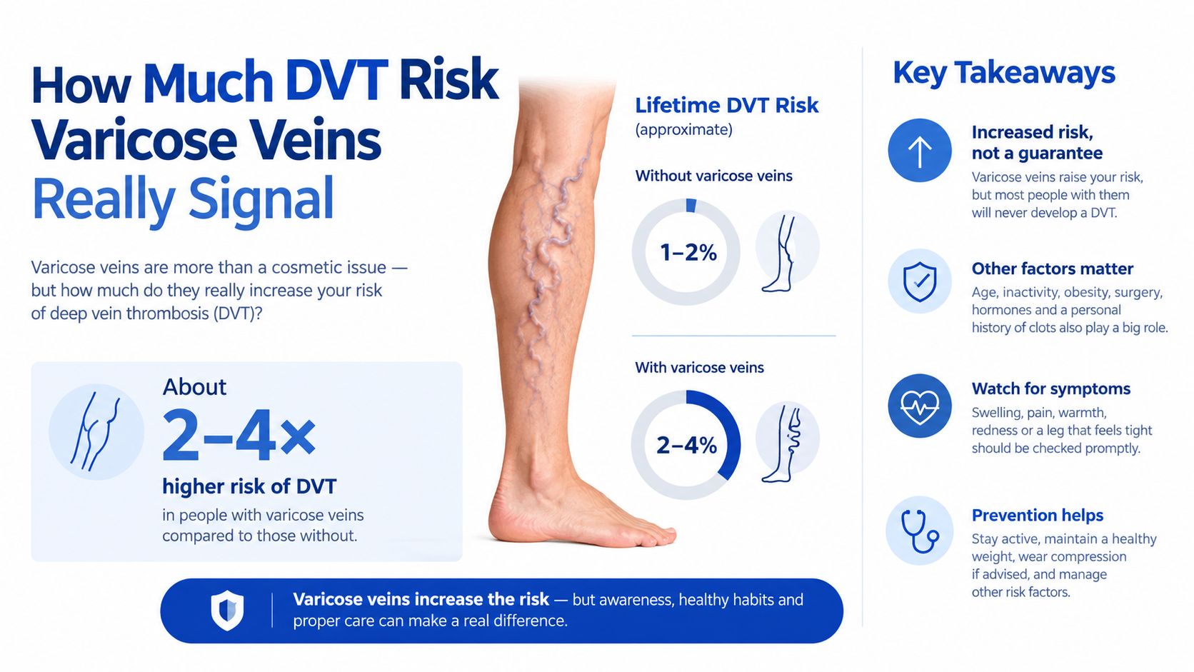
Varicose veins are not just a surface problem. Cohort evidence has shown that they can mark a meaningfully increased DVT risk profile, including a hazard ratio of 5.3 in one Taiwan health-insurance analysis and a U.S. population-based study in which DVT incidence was 6.55 per 1,000 person-years in patients with varicose veins versus 1.23 per 1,000 person-years in controls, with an absolute risk difference of 5.32 per 1,000 person-years (JAMA study). A large Taiwanese cohort of 425,968 adults also found higher DVT risk and higher PE risk in patients with varicose veins (cohort review).
Relative risk and absolute risk are not the same thing
Relative risk makes the association look dramatic. Absolute risk keeps it grounded. For most patients, visible varicosities are a risk marker, not a diagnosis of clotting disease. That distinction matters because the right response is usually not panic, it's a lower threshold for duplex ultrasound when symptoms change.
The patients I worry about most are the ones with prior clot, active malignancy, recent hospitalization, known thrombophilia, or unilateral symptomatic varicosities that suddenly become more painful or swollen. Those features push the leg out of the routine category and into the image-now category.
If varicose veins are stable, the concern is usually symptom control and chronic venous management. If they become suddenly painful, warm, or asymmetric, the concern shifts to thrombosis until imaging says otherwise.
Diagnosis and When to Get Imaging
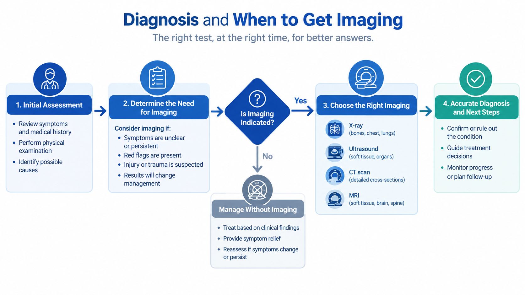
Duplex ultrasound is the shared backbone here. In suspected DVT, compression duplex ultrasound checks whether a vein compresses normally. In varicose veins, the same exam shows reflux, vein diameter, valve competence, and superficial flow patterns in one study, which is why the internal ultrasound and treatment resource is useful for scheduled vein assessment.
When the scan should happen now
Same-day imaging is reasonable when symptoms start suddenly and affect one leg. New swelling with pain, calf pain after immobilization, sudden discoloration, hardening along a varicose segment, or new symptoms in a patient with known malignancy all justify a lower threshold for urgent duplex ultrasound.
D-dimer can help in low-probability DVT workups, but it is less useful after recent surgery, during pregnancy, or when inflammation is already active.
If the question is how clinicians think about clot detection across imaging methods, finding blood clots with Lola is a useful general reference point.
Chronic heaviness, bulging veins, ankle swelling that builds through the day, and skin changes near the ankle still deserve evaluation. They usually point to outpatient vein workup rather than an emergency clot assessment, especially when the goal is to document reflux and plan treatment rather than rule out an acute clot. The internal ultrasound and treatment resource fits that setting.
Treatment Paths for Each Condition
Treatment should match the disease process in front of you. Varicose veins call for symptom relief, reflux reduction, and skin protection. DVT calls for clot prevention, clot treatment, and limiting long-term venous damage. Those goals do not overlap, so treating one does nothing for the other.
| Treatment Category | Varicose Veins | Deep Vein Thrombosis |
|---|---|---|
| First step | Compression, elevation, activity, weight control | Anticoagulation and mobilization guidance |
| Procedure options | Endovenous thermal ablation, foam sclerotherapy, mechanochemical ablation, microphlebectomy, limited stripping | Catheter-directed thrombolysis or thrombectomy in selected iliofemoral cases |
| Medication role | Symptom support, not clot treatment | Core therapy, often with DOACs or warfarin with bridging |
| Main goal | Relieve symptoms, protect skin, reduce reflux | Prevent clot propagation and pulmonary embolism |
What works for reflux versus what works for clot
For varicose veins, conservative care usually starts with graduated compression, leg elevation, activity, and weight control. If reflux still causes symptoms, office-based treatment can include thermal ablation, ultrasound-guided foam sclerotherapy, mechanochemical ablation, and microphlebectomy. For DVT, the main step is anticoagulation, often for a finite treatment course, with compression and ambulation used to support recovery and limit post-thrombotic symptoms.
A practical detail matters here. A superficial clot near the deep venous junction is not a minor vein problem. Proximity changes the risk profile, so anticoagulation may be needed. That is one more reason visible veins and clot risk should be assessed together.
Vein and wound care can also be handled in one setting, and some practices, including Vein and Wound Institute, combine duplex-based evaluation with minimally invasive vein treatment and wound-focused follow-up when chronic venous disease and skin breakdown overlap. The point is coordination. The reflux problem and the tissue problem are addressed together.
The main treatment error is trying to make varicose vein care do the job of DVT care, or the reverse. One is elective reflux management, the other is acute clot management.
When to Seek Urgent Care Versus a Vein Evaluation
A useful triage framework has three tiers. Red flags mean emergency evaluation. Yellow flags mean a prompt vein workup. Green flags usually mean routine follow-up or self-management with compression if it's already been recommended.
Red, yellow, and green triage
Red-flag symptoms include sudden unilateral calf or thigh swelling, severe pain with dorsiflexion, chest pain or shortness of breath suggesting pulmonary embolism, a cord-like tender vein with redness extending above the knee, and any leg swelling after recent immobilization, surgery, or active cancer. These are not “watch it for a few days” symptoms.
Yellow-flag findings are new visible varicose veins with aching, heaviness, night cramps, ankle edema that worsens during the day, skin discoloration near the ankle, a healed or active venous ulcer, prior superficial thrombophlebitis, or a strong family history of venous disease. These deserve a scheduled evaluation because they suggest chronic venous disease that may be progressing.
Green-tier patterns include long-standing asymptomatic varicose veins, mild cosmetic concerns, and routine compression wear without new symptoms. Those cases still deserve attention if the appearance or symptoms change, but they usually don't require urgent imaging.
| Triage Tier | What It Looks Like | Action |
|---|---|---|
| Red | Sudden unilateral swelling, severe pain, dyspnea, chest pain | Emergency department |
| Yellow | Chronic heaviness, skin change, ulcer, prior superficial clot | Vein evaluation within days to weeks |
| Green | Stable, long-standing, minimally symptomatic veins | Routine follow-up |
When in doubt, duplex ultrasound within 24 to 48 hours is a reasonable middle ground for uncertain but non-crashing symptoms. Sudden unilateral pain, swelling, or shortness of breath belongs in the emergency department, not a routine vein visit.
If you're sorting out whether your symptoms fit chronic reflux or a possible clot, get a proper venous exam instead of guessing from appearance alone. The Vein and Wound Institute evaluates venous disease, uses duplex ultrasound to clarify the cause, and treats chronic varicose-vein problems and wound-related complications in one coordinated setting. Visit Vein and Wound Institute to arrange an evaluation if your leg symptoms are new, one-sided, or getting worse.